Using an off-the-shelf digital camera, Rice University biomedical engineers and researchers from the University of Texas M. D. Anderson Cancer Center have created an inexpensive device that is powerful enough to distinguish oral cancer cells from healthy cells simply by viewing the LCD monitor on the back of the camera.
The results of the first tests of the camera were published online June 23 in the open-access journal PLoS ONE.
"Consumer-grade cameras can serve as powerful platforms for diagnostic imaging," said Rebecca Richards-Kortum, Ph.D., a professor of bioengineering at Rice and the study's lead author. "Based on portability, performance, and cost, you could make a case for using them both to lower healthcare costs in developed countries and to provide services that simply aren't available in resource-poor countries."
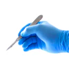
In this study, Richards-Kortum and her team captured images of cells with a small bundle of fiber-optic cables attached to a $400 Olympus E-330 camera. A human tissue specimen was imaged following surgical resection, enabling dysplastic and cancerous regions to be evaluated. The oral mucosa of a healthy human subject was imaged in vivo, following topical application of 0.01% proflavine.
The proflavine caused cell nuclei in the samples to glow brightly when lighted with the tip of the fiber-optic bundle. The researchers tested three tissue types: cancer cell cultures that were grown in a lab, tissue samples from newly resected tumors, and healthy tissue viewed in patients' mouths.
 |
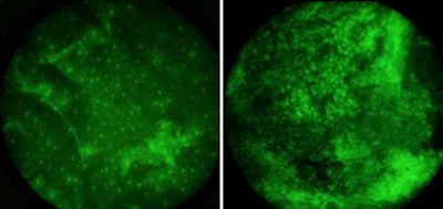
| The green spots in these images are cell nuclei treated with a fluorescent dye. The cells are from the oral cavity of cancer patients. In healthy tissue (left), the nuclei are small and widely spaced. The nuclei of cancer cells (right) are abnormally large and close together. Image courtesy of Rice University. |
Because the nuclei of cancerous and precancerous cells are notably distorted from those of healthy cells, abnormal cells were easily identifiable, even on the camera's small LCD screen, Richards-Kortum said.
"The dyes and visual techniques that we used are the same sort that pathologists have used for many years to distinguish healthy cells from cancerous cells in biopsied tissue," said study co-author Mark Pierce, Rice faculty fellow in bioengineering. "But the tip of the imaging cable is small and rests lightly against the inside the cheek, so the procedure is considerably less painful than a biopsy and the results are available in seconds instead of days."
Co-authors of the paper include Dongsuk Shin and Mark Pierce, Ph.D., both of Rice, and Ann Gillenwater, M.D., and Michelle Williams, M.D., both of the University of Texas M. D. Anderson Cancer Center. The research was funded by the National Institutes of Health.
Copyright © 2010 DrBicuspid.com