Clinicians should suspect underlying dental conditions when patients present with a rare, potentially life-threatening Pott's puffy tumor or frontal bone osteomyelitis, according to a review published in the February issue of the Journal of Oral and Maxillofacial Surgery.
Patients who have a Pott's puffy tumor should receive comprehensive oral cavity and dental examinations and should be referred to dental colleagues when necessary, as initial imaging may not identify all dental disease, the authors advised (J Oral Maxillofac Surg, February 2021, Vol. 79:2, pp. 389-397).
"In the absence of a history of frontal bone trauma or frontal sinus surgery, underlying dental origin should be suspected in cases of PPT," wrote the group, led by Dr. Rohit Nallani from the department of otolaryngology-head and neck surgery at the University of Kansas Medical Center in Kansas City.
Pott's puffy tumor presents with soft swelling over the frontal bone and can lead to compression of structures and neurological, gastrointestinal, and other symptoms. The condition can sometimes lead to serious diseases such as meningitis, venous sinus thrombosis, or sepsis.
Pott's puffy tumor is a rare side effect of bacterial sinusitis or complication of facial trauma. However, some research has shown that it may occur due to facial plastic surgery, intranasal drug abuse, and odontogenic disease. The researchers aimed to better understand the relationship between concurrent dental disease and PPT, especially when patients have no history of facial trauma or prior frontal sinus surgery, they wrote.
Nallani and colleagues conducted a retrospective chart review of patients diagnosed with PPT between 2010 and 2019; 17 patients were identified. They analyzed the patients' demographics, medical history, procedures, microbial cultures, and antibiotics taken. Then, maxillofacial computed tomography (CT) scans were reviewed to determine which patients had odontogenic disease.
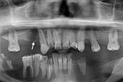
Among the patients diagnosed with PPT, 16 (94%) had odontogenic disease visible on CT scans, and 12 (75%) of the 16 had no previous frontal sinus trauma or surgery. Initial radiology reports cited odontogenic disease in seven patients (41%), but the CT scans showed various pathology in 16 patients upon dentist review.
Despite the findings, there were some study limitations, such as the data being collected from patient charts. In the clinical and operative notes left by physicians, especially when patients saw multiple doctors, there was variability in the description of findings and procedure terminology.
Clinicians encountering patients with this rare condition should evaluate the oral cavity and teeth, request further imaging, and refer them to other professionals as needed.
"In patients without a previous history of frontal sinus surgery or bony trauma, odontogenic disease, especially of the maxillary teeth, may be a contributing factor to frontal bone osteomyelitis," they wrote.