Imaging aided in the treatment of a woman who experienced months of nasal congestion and blood-tinged secretions due to a rare intranasal tooth thought initially to be a rhinolith. The case report was published on February 28 in the Cureus Journal of Medical Science.
A rigid nasal endoscopy revealed what appeared to be a rhinolith on the floor of the left nasal cavity. Clinicians determined it was a tooth because it had enamel and cementum.
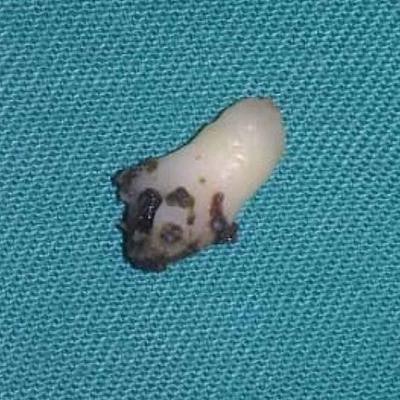
 The tooth retrieved from the nasal cavity. All images courtesy of Ong et al. Licensed under CC BY-NC 4.0.
"Detailed dental and oropharyngeal examination as well as imaging studies are essential in diagnosing an intranasal tooth," wrote the authors, led by Dr. Hui Yan Ong from the University of Malaya Medical Centre in Kuala Lumpur, Malaysia.
Though the presence of a tooth in the nasal cavity is uncommon, it occurs more often in patients with a cleft lip or palate. The incidence of an intranasal tooth in the general population is between 0.1% and 1%. The exact mechanism is unknown but may also include crowded teeth leading to eruption upward or the occurrence of orofacial trauma.
 An endoscopic view of the patient's left nasal cavity. There was a hard, whitish mass (blue arrow) at the floor of the cavity.
A 24-year-old woman
For six months, the patient experienced nasal obstruction in her left nostril and intermittent blood-stained discharge that was unprovoked. Her history was unremarkable, other than having undergone cleft palate repair earlier in life. She had no other symptoms and no history of rhinitis; however, her primary care doctor treated her for rhinitis. She felt little relief after using steroidal nasal spray and antihistamines, so she was referred to an otolaryngologist for further evaluation.
During an exam, the otolaryngologist noticed a small deformity over her left incisor area, which appeared to be misaligned. A cold spatula test revealed her to have reduced nasal flow in the left nostril compared to the right. When a rigid nasal endoscopy was performed, it revealed what appeared to be a rhinolith, or nasal stone, on the floor of the left nasal cavity.
 Part of the maxillary arch; canines and the left upper premolar teeth are well formed. There are two malformed incisors between the canines.
The examination and removal of the rhinolith, which turned out to be a tooth, were done under general anesthesia. She was discharged after surgery and no longer had any nasal symptoms, the authors wrote.
Early treatment, better outcomes
Early surgical removal of an intranasal tooth is necessary to prevent more serious complications. If patients do not respond to conventional treatments, they should undergo ear, nose, and throat and endoscopic evaluations, they wrote.
 An endoscopic view of the woman's nasal cavity after tooth removal. The nasal cavity floor appeared intact (blue arrow).
"Intranasal tooth is uncommon; however, it should be one of the differential diagnoses to be considered for patients presenting with unilateral nasal symptoms," the group concluded.