High-resolution magnetic resonance imaging (MRI) can effectively visualize variations in the inferior alveolar nerve of the mandible, and should be used in vague or unclear oral surgery cases when orthopantomography and CT scans will not suffice, according to a study in the Journal of Computer Assisted Tomography (January/February 2012, Vol. 36:1, pp. 150-153).
"Anatomical variations of the dental canal structures are present in a small proportion of the population and often go undiagnosed," wrote the study authors, from Aachen University. "In such cases, there could be an increased risk of complication during surgery and failure of anesthesia."
Preoperative knowledge of the position of neural structures can help avoid complications such as injury to the neurovascular bundle, they noted. Damage to these nerves during a clinical intervention can be severe and can include impairment, paresthesia, and pain. Such injuries could also have legal consequences, according to the researchers.
Computed tomography and other forms of digital volume tomography provide good resolution and contrast between soft tissues and bones, and are relatively inexpensive imaging modalities. However, "these diagnostic methods are not always suitable for exact delineation of the neural structures in the mandible," the study authors wrote, adding that "the exact knowledge of precise anatomical structures, and especially because of rare variation in the mandible, is an essential foundation to detect pathological processes in diagnosis and planning of surgery."
Orthopantomography is routinely used for presurgical planning but has the disadvantage of distortion, the researchers noted. CT scans, meanwhile, expose the patient to much higher radiation doses, and dental materials can cause artifacts on these scans.
MRI, on the other hand, can delineate soft tissue and bone marrow much more precisely, which provides a good foundation for discriminating fine anatomic details. In addition, MRI does not expose the patient to ionizing radiation.
Unique variations found
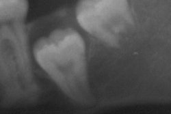
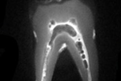
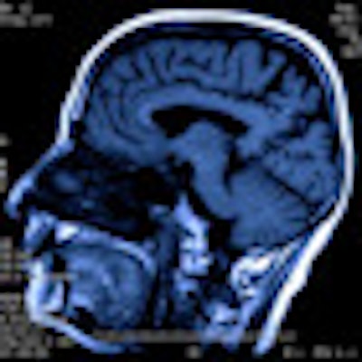
To demonstrate MRI's ability to obtain detailed information about canal structures in the mandible, the researchers had 64 patients (39 women, 25 men) undergo a routine MRI exam of the head and neck. The exams were performed with a 3-tesla Magnetom Verio MRI system (Siemens Healthcare) in axial orientation parallel to the occlusal plane.
Two radiologists analyzed the images with respect to anatomical features, variations, and rarities of the mandibular canal, mental canal, incisive canal, and nutrient canals, and noted the frequency of variations. They found a total of 16 variations in 25% of the patients.
The variations included the following:
- Winding mandibular canal (2)
- Double mandibular canal (2)
- Double incisive canal (5)
- Branch out of mental canal (3)
- Medial incisor supplied by the other side (1)
Four of these variations -- the winding canal, the mandibular canal running laterally to the teeth, the canal with large caudolingual branches, and the duplicated incisive canal in the anterior chin region -- have not been previously reported in the literature, according to the study authors.
This study is a nice example of why dentists and dental specialists need MRI, Allan Farman, BDS, MBA, PhD, DSc, a professor of radiology and imaging science at the University of Louisville in Kentucky, told DrBicuspid.com.
"One of the American Academy of Oral and Maxillofacial Radiology requests is going to be to add maxillofacial MRI to the CDT [Code on Dental Procedures and Nomenclature] codes for the first time," he said. "Most of the advanced imaging conducted by specialists in oral and maxillofacial radiology are not covered by the CDT codes at this point in time. Indeed, most of the CDT codes for radiography still specify 'film,' while the world of dentistry has gone largely digital."
While MRI is not a routinely used imaging modality in dentistry at this time, "there are some major advantages, as we demonstrated in this study," the study authors wrote.
The application of MRI technologies in dentistry and oral surgery can improve patient care, and MRI should be performed in vague or unclear cases that could not be clarified by orthopantomographs or CT scans.