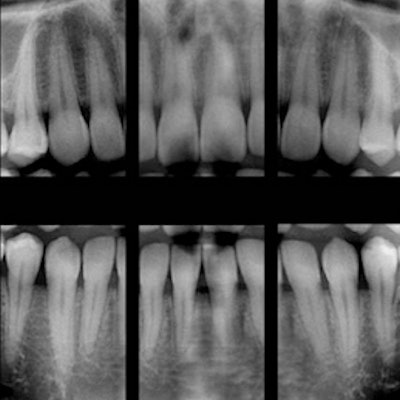
The usefulness of bitewing images for helping find caries in permanent teeth is well-established. But is the modality also useful in determining caries in primary teeth? Researchers compared the images to visual examination to find out.
They found more than 400 additional caries in just over 500 children when studying the images compared with a visual examination of the child's mouth. Their study findings were published in BMC Oral Health on August 9.
"Diagnostic yields from bitewing radiographs are greater for children with greater caries experience," L. A. Foster Page, BDS, PhD, and colleagues wrote. Dr. Page is in the department of oral sciences at the University of Otago Faculty of Dentistry in Dunedin, New Zealand.
Primary query
The use of bitewing radiographs has been shown to help in the estimation of caries in permanent teeth, but there is a lack of clinical information on their use in primary teeth, particularly molars. The researchers wanted to see how bitewing radiography compared with clinical examination in detecting caries in primary molars.
“Diagnostic yields from bitewing radiographs are greater for children with greater caries experience.”
They examined more than 500 children ages 3 to 8 in New Zealand. Almost 75% of those examined were younger than 6 years old. Each child underwent a clinical examination by one of 13 dental therapists, who recorded the clinical status of the patient's primary dentition. A carious lesion was counted if cavitation was present. Researchers used the number of decayed, missing, and filled surfaces (dmfs) in the primary dentition to separate the children into one of three groups: 0 dmfs, 1 to 8 dmfs, or 9+ dmfs.
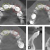
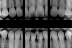
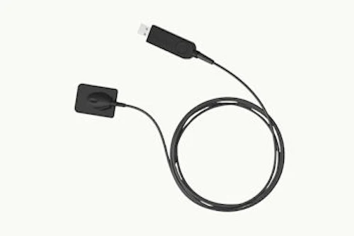
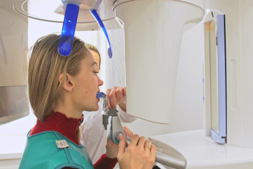
Bitewing radiographs also were taken at the time of the clinical examination. A Belmont Belray 096-C (Belmont) system was used for the images.
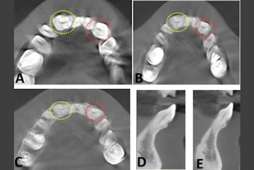
After clinical examination, caries prevalence was 63.1% and mean dmfs was 4.6. After radiographs, the prevalence was 74.7% and mean dmfs was 5.8. In the 185 children with no apparent caries at clinical examination, 124 lesions were detected radiographically (see table below).
| Comparison of caries found on clinical examination vs. bitewing radiographs | ||
| dmfs | No. of caries with clinical examination | No. with bitewings |
| dmfs = 0 (n = 185) | 0 | 124 |
| dmfs = 1-8 (n = 218) | 342 | 556 |
| dmfs = 9 or more (n = 98) | 308 | 414 |
| Overall (n = 501) | 650 | 1,094 |
Worth the challenge
While the researchers did not list any limitations, they noted that the study did not include caries lesions limited to the enamel, so the findings may have underestimated the true disease level.
They also cautioned that the focus of this study was the lesions themselves rather than their prevention or restorative treatment. They clearly stated that they were not necessarily advocating restoration of all the extra lesions discovered, noting that was a clinical decision to be made by the treating clinician.
The researchers concluded that, while taking bitewing radiographs in young children is not without challenges or risks, the results of their study backed their use.
"The findings of this study further support the need to consider using bitewing radiographs in young children to enhance the management of lesions not detected by a simple visual examination alone," the authors wrote.