Dear Imaging & CAD/CAM Insider,
Three-dimensional imaging has become a standard tool for implant diagnostics and surgical treatment planning. But it has the potential to play a much larger role, particularly in periodontics, according to two pioneers in the field.
In the latest installment of our Leaders in Dentistry series, we spoke with Dr. Alan Rosenfeld and Dr. George Mandelaris about their recent experience working with cone-beam CT in their periodontal practice. They believe this technology puts periodontists in an even more critical position to enable correct diagnoses and assign more accurate prognoses, which in turn can enhance treatment planning for interdisciplinary dental teams.
Click here to read more in this Imaging & CAD/CAM Insider Exclusive.
In related news, a European consortium has developed comprehensive guidelines for the appropriate use of cone-beam CT in dentistry, including referral criteria, radiation protection of patients and staff, training, and quality assurance procedures. Can the U.S. be far behind?
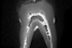
And magnetic resonance imaging (MRI) continues to attract the attention of dental researchers around the world. Several studies published in recent months showcased the potential for MRI as a hard- and soft-tissue diagnostic tool in dentistry, with research groups in Germany leading the way. Read more.
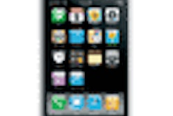
Meanwhile, smartphone apps have become all the rage, and dentistry is no exception. While many of the current dental apps are educational, a Canadian start-up has developed a smartphone tool that better connects dental practices with dental labs and streamlines the prescription-writing process.
In other Imaging & CAD/CAM Community news, optical coherence tomography (OCT) is a noninvasive approach for localizing the most representative site to biopsy in a suspicious oral lesion, according to a study in Lasers in Medical Science. And OCT combined with polarimetry yields a powerful tool for detecting and diagnosing the dysplasia that typically precedes oral squamous cell carcinoma, according to a study in the Journal of Biomedical Optics.
Meanwhile, most U.S. states require a periodic assessment of all radiographic equipment in a dental, medical, or hospital setting. Now a novel device for evaluating the image quality and patient dose of intraoral x-ray systems is gaining favor among radiation protection agencies and dental offices.
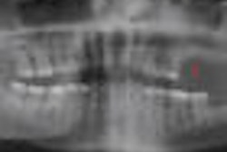
And finally, certain commonly used image compression techniques -- notably JPEG -- can affect the diagnostic quality of panoramic images and the ability to evaluate certain anatomical structures in these images, according to a study in Clinical Oral Investigations. Read more.