A dentin bridge or a layer of hard dental tissue formed spontaneously underneath a 54-year-old man’s tooth with pulp necrosis and internal resorption, which is highly unusual. The case report was published recently in Radiology Case Reports.
Cone-beam computed tomography (CBCT) revealed that the dentin bridge preserved the vitality of the tooth’s apical pulp. The man underwent a root canal limited to the necrotic coronal pulp up to the dentin bridge and restoration with a composite. A follow-up x-ray at three months showed treatment stability, the authors wrote.
“Spontaneous dentin bridge formation in mature teeth without pulp exposure or therapeutic intervention is an exceedingly rare phenomenon,” wrote the authors, led by Dr. Bita Heydarzadeh of the Department of Oral and Maxillofacial Radiology, School of Dentistry, at the Shahid Beheshti University of Medical Sciences in Iran (Radiol Case Rep, January 5, 2026, Vol. 21:3, pp. 1346-1351).
A 54-year-old man with a gray, discolored incisor
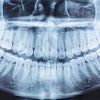
The man had tooth discoloration after dental trauma. An exam revealed coronal pulp necrosis, and periapical radiography and CBCT confirmed internal resorption in the coronal pulp chamber with a spontaneously formed dentin bridge, the authors wrote.
 Fig. 1: Initial images.Images and captions courtesy of Heydarzadeh et al. Licensed under CC BY 4.0.
The dentin bridge appeared to separate the necrotic coronal pulp from vital tissue. This thick barrier likely added to the preservation of apical pulp vitality, which was proven by the lack of apical pathosis and a normal apical canal configuration as shown on the CBCT, they wrote.
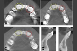
 Figure 2: Occlusal views of CBCT images showing internal root resorption, bone loss, and perforation of the cortical plates.
 Figure 3: Coronal view of a CBCT image shows the dentin bridge separating the necrotic coronal pulp from the vital apical pulp.
To treat the patient, he underwent root canal therapy limited to the necrotic coronal pulp up to the dentin bridge. Internal bleaching followed to improve the discoloration, and then a restoration with composite resin was completed.
At his three-month follow-up, a radiograph showed no problems, according to the case report.
 Figure 4: Photo and periapical radiography after root canal therapy and tooth restoration.
What was learned
This case report revealed that a spontaneous dentin bridge, produced by odontoblast-like cells, can preserve apical vitality in traumatized teeth. Additionally, CBCT was necessary for diagnosis and treatment planning, and conservative root canal treatment up to the dentin bridge is achievable, the authors wrote.
However, the case report had limitations, including that odontoblastic activity was not histologically confirmed, they wrote.
“This case demonstrates rare spontaneous dentin bridge formation post-trauma, enabling conservative treatment,” Heydarzadeh and colleagues wrote.