Scientists from multiple institutions are launching a research effort to identify cellular principles that guide how facial bones take shape.
Led by Kristen Prufrock, PhD, from Washington University in St. Louis, the team recently received a $600,000 grant from the National Science Foundation. The funds will be used to develop 3D reconstructions of dental and bone features throughout the developmental stages. Prufrock hopes that the reconstruction models will help explain the role of developing dentition in shaping the facial form.
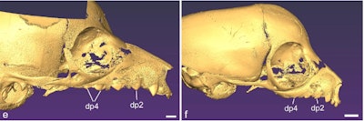
The researchers will evaluate samples from multiple species of lemurs and monkeys. The primates vary in regard to the pace of tooth development and snout length. The team will use high-resolution micro-CT imaging to visualize and quantify soft tissue in a 3D fashion. Other researchers will label tissue with markers for spatial patterns of cellular activity along the perimeter of tooth germs as it relates to ossification of the developing jaw.
Ultimately, the goal of the study is to better understand the cellular basis for the functional matrix theory, where bones take direction from the soft tissue and spaces around them during development. The group added that it will freely share its models for research and teaching purposes.