A new software tool used in conjunction with an intraoral camera demonstrated high sensitivity in the automatic, real-time detection of root canal orifices, according to a study in the Journal of Dental Education (November 2011, Vol. 75:11, pp. 1452-1457).
Researchers from University Medical Center-Mainz and University Medical Center of the Johannes-Gutenberg University previously demonstrated the software's ability to detect root canal orifices using a video camera mounted on a microscope (Journal of Endodontics, October 2009, Vol. 35:10, pp. 1400-1403).
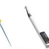
For this study, the researchers used a Durr Dental VistaCam Digital intraoral camera system connected to an HP Compaq nc8430 laptop with 2-GHz processor and 2 GB of RAM to study 200 human molars with fully intact root and enamel surfaces and without endodontic treatments. No third molars were included, and only teeth recently extracted for periodontal reasons were included.
"The tested software is able to scan the video stream of an intraoral camera connected for the coronal contours of root canal orifices according to the method described in an earlier study," the researchers wrote. "The user can place the camera above the access cavity of the desired tooth, and detection can be activated via the mouse button."
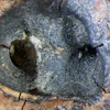
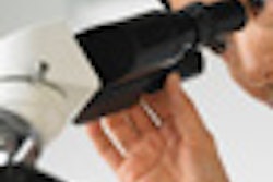
The teeth were positioned beneath the intraoral camera so that the entire pulp canal floor and parts of the crown were visible. After the first series of 200 digital images was taken, an experienced observer (with five years of experience in endodontic treatment) evaluated the images separately on the computer screen. The same images were then evaluated by a predoctoral student.
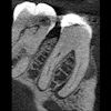
At this point, the researchers implemented the new software tool, taking real-time video images of each tooth using the intraoral camera and storing screenshots of the detection results for subsequent analysis by the observer and statistical evaluation. Histological samples also were then prepared for comparison.
High sensitivity
The histological examination of all teeth revealed 529 root canals. The experienced observer marked 503 root canal orifices in the images, of which 479 were verified when compared with the histological slices and the other 24 were false positives (90.6% sensitivity).
The predoctoral student marked 455 root canal orifices, of which 448 were verified and the remaining seven found to be false positives (84.7% sensitivity).
The software found 543 root canal orifices, of which 506 were verified by the histological standard and 37 were found to be false positives (95.7% sensitivity).
"The results for the sensitivity of the software compared with histological slices are very promising for a possible computer-assisted application," the study authors wrote.
The most remarkable differences were found for the detection rate for the second mesiobuccal canal orifices, they added. The experienced observer detected 42.5% of the orifices in the second mesiobuccal canals, while the predoctoral student detected 8.7% and the software detected 60.9%.
Based on these findings, this software could be a useful computer-assisted endodontics educational tool for pre- and postdoctoral students and warrant in vivo testing of the system, according to the researchers.
"Our study indicates that the software is capable of automatic real-time detection of root canal orifices with intraoral cameras," they wrote. "The tested software reaches a high sensitivity for automatic real-time detection of root canal orifices with intraoral cameras in direct comparison to histological images."
The software could also be used to collect statistical data on root canal orifice shapes, distances, and other anatomical features of interest, they added.
"The idea is to collect anatomical tables from ideal teeth, save them into a database, and use them for a future augmented reality system for computer-assisted learning or diagnosis," the authors concluded.