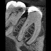
Figure 1: Cone-beam computed tomography image, sagittal view, tooth #30.Images courtesy of Dr. Juan F. Yepes.
DrBicuspid publishes a new case study weekly. To test your dental expertise, first, please log in. If you don't have a login, create one. Each case comprises a history, quiz, and discussion section. An answer key is provided at the end of the case study.
A 40-year-old woman was referred to the endodontist by her family dentist to evaluate a lesion located at the periapical area of tooth #30. The woman was asymptomatic. She did not report paresthesia, and there was no history of trauma.
Log in to view the full article
Figure 1: Cone-beam computed tomography image, sagittal view, tooth #30.Images courtesy of Dr. Juan F. Yepes.
DrBicuspid publishes a new case study weekly. To test your dental expertise, first, please log in. If you don't have a login, create one. Each case comprises a history, quiz, and discussion section. An answer key is provided at the end of the case study.
History
A 40-year-old woman was referred to the endodontist by her family dentist to evaluate a lesion located at the periapical area of tooth #30. The woman was asymptomatic. She did not report paresthesia, and there was no history of trauma.
Her past medical history included seasonal allergies and a recent COVID-19 infection.
The extraoral and intraoral exams were within normal limits. There was no tooth mobility. The endodontist ordered a cone-beam computed tomography (CBCT) scan. Below is a sagittal and axial view of the tooth.
Figure 1: Cone-beam computed tomography image, sagittal view, tooth #30.Images courtesy of Dr. Juan F. Yepes.
 Figure 2: CBCT, axial view, tooth #30.
The endodontist consulted with an oral and maxillofacial radiologist.
What is the LEAST likely preliminary diagnosis?
A. Unicystic ameloblastoma
B. Odontogenic keratocyst
C. Dentigerous cyst
D. Radicular cyst
E. Idiopathic bone cavity
2. The oral radiologist evaluated the CBCT films and provided a report to the endodontist. A paragraph from the report is provided below:
"Small hypodense lesions (radiolucent) are observed at the mesial and distal apices of tooth #30. The lesions are well defined and well corticated. There is no evidence of expansion in the coronal and axial projections for the small lesion at the distal apex. However, there is evidence of expansion (buccal) of the hypodense lesion located at the apex of the mesial root. Hyperdense bone is present at the periphery of the lesion, indicating a chronic inflammatory process."
Based on the report, what is the most likely diagnosis from the radiologist?
A. Ameloblastoma
B. Odontogenic keratocyst
C. Cemento-osseous dysplasia
D. Radicular cyst
E. Fibrous dysplasia
3. Which of the following is a critical step to perform in determining a precise differential diagnosis?
A. Test the vitality of the teeth associated with the lesion.
B. Check for the presence of symptoms associated with the lesion.
C. Inquire about the patient’s family dental history.
D. A and B are correct.
E. A, B, and C are correct.
4. The endodontist performed an excisional biopsy and a root canal treatment. The histopathology report confirmed the diagnosis of a radicular cyst.
Which of the following is true about the origin of radicular cysts?
A. They occur as a result of reduced enamel epithelium around the apex of a vital tooth
B. They are the remnants of epithelial cells around the apex of a nonvital tooth
C. They originate from periodontal ligament cells around the apex of a vital tooth
D. None of the above
5. Radicular cysts belong to the odontogenic inflammatory cyst group.
A. True
B. False
6. Where specifically do radicular cysts originate from?
A. Epithelial remnants of Malassez
B. Epithelial cells around the dental follicle
C. Epithelial cells around the cemento-enamel junction
D. B and C are correct
Discussion
Radicular cysts are the most common odontogenic inflammatory cyst of the jaws. They comprise about 50% of all cysts that can affect the maxilla and the mandible. They are typically asymptomatic unless they are infected.
Radicular cysts originate on epithelial remnants (Malassez), which are stimulated and proliferate by an inflammatory process from a nonvital tooth. The cyst originates from a necrotic pulp which remains in situ long enough to develop a chronic inflammatory process. The most common site is the apex of a nonvital tooth, quite often the anterior maxillary teeth.
From a radiographic perspective, radicular cysts are completely radiolucent (in long-standing radicular cysts, calcifications are sometimes present), well defined, and well corticated. Resorption or displacement can be associated with the cyst. Depending on the size of the lesion and the feasibility of restoring the tooth, treatment options can range from root canal treatment to conservative surgical removal. The prognosis is excellent.
References
1. Neville BW, Damm DD, Allen CM, Bouquot JE. Oral and Maxillofacial Pathology. 3rd ed. St. Louis, MO: Saunders/Elsevier; 2009.
2. White SC, Pharoah MJ. Oral Radiology: Principles and Interpretation. 6th ed. St. Louis, MO: Mosby; 2009.
Note: Dr. Yepes would like to thank Dr. Zane Lambert, Terre Haute, IN, for sharing this case.
Answer key
C; dentigerous cyst.
D; radicular cyst.
D; A and B are correct.
B; They are remnants of epithelial cells around the apex of a nonvital tooth.
A; true.
A; epithelial remnants of Malassez.