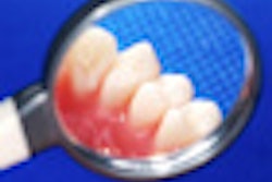
It seems there's no escaping the need to assess clinical attachment loss (CAL) in at least some teeth of patients undergoing periodontal treatment. A study published in the Journal of the American Dental Association has confirmed that measuring probing depth (PD) alone is unreliable in all but the most severe periodontitis cases for determining whether the disease is progressing or not (February 2013, Vol. 144:2, pp. 171-178).
 Bryan Michalowicz, DDS, MS.
"PD leads to underestimation of CAL when there is gingival recession," wrote the study authors, from the University of Minnesota. "Conversely, PD leads to overestimation of CAL when the gingiva is enlarged because of, for example, inflammation or use of certain medications."
Determining the CAL at any given location takes about twice as long as determing the PD, they noted. CAL is calculated by measuring both the PD and the distance from the cementoenamel junction (CEJ) to the gingival margin (GM). Previous research has shown that measuring PD alone does not reliably reflect whether teeth have progressive CAL, except in sites with deep PD to begin with, the researchers noted.
To confirm and extend these findings, Bryan Michalowicz, DDS, MS, a professor and the Erwin Schaffer Chair in Periodontal Research in the department of developmental and surgical sciences, and two colleagues reviewed information from 314 patients who had been involved in clinical trials of nonsurgical periodontal treatment at the center and who had at least 12 months of follow-up data.
To determine whether it is better to measure the CAL and PD more often than once a year, and/or to only monitor CAL at sites that show a PD increase of at least 2 mm, the team compared the following regimens that were applied at baseline and one year later:
- Monitoring PD at six sites on all teeth
- Monitoring PD at six sites/teeth and CAL at the midfacial and midlingual sites only
- Assessing PD and CAL at six sites/teeth at baseline, monitoring PD at six sites/teeth, and then measuring CAL only at sites that have an increase in PD of at least 2 mm
Comparing apples to apples
At baseline, 47.4% of the tooth sites had a PD of at least 5 mm and 67.4% had a CAL of at least 3 mm. After the treatments in the clinical trials, more teeth were in better periodontal condition according to the PD and CAL assessments than had worse disease.
“It may be reasonable to monitor PD alone in patients with a thick gingiva and bone around the teeth.”
of Minnesota
The second option -- monitoring PD at six sites/teeth and CAL at the midfacial and midlingual sites only -- had a sensitivity of 54.4%, compared with 32.4% for the other two. The specificities, positive predictive values (PPVs), and negative predictive values (NPVs) were high for all three options, with the third option hitting a specificity and PPV of 100% and an NPV of 95.9%.
However, changes in PD were not highly correlated with changes in CAL. The researchers found that a change in PD of at least 2 mm on the mesial or distal surfaces had a sensitivity of 49% for predicting a CAL loss of at least 2 mm. The sensitivity for PD at the molars was similar, at 48%, while it was 38% at the nonmolars and 24% at the midfacial or lingual surfaces.
Moreover, the smaller the PD, the weaker the association, with sensitivities of only 18% for PDs of 3 mm or less and 74% for PDs of at least 7 mm. This is despite the fact that the PPVs were high: Approximately 70% of sites with a PD change also had a CAL change in the same direction.
The results weren't any more promising when the team focused only on teeth with progressing periodontitis. The PPVs and NPVs were reasonably robust, but the sensitivities ranged from 24% for midfacial or lingual sites to 41% for molars, and from 29% for PDs of 3 mm or less to 47% for PDs of at least 7 mm.
"We found that use of PD alone to assess changes in a patient's periodontal status frequently fails to detect changes in CAL, especially at initially shallow and moderate sites. Clinicians should consider monitoring the position of the GM relative to the CEJ, which is used to compute CAL, in patients undergoing periodontal treatment," the study authors concluded.
CALmonitoring may be most meaningful in patients with thin facial or lingual tissues, according to Dr. Michalowicz.
"In these patients, progressive CAL may be detected on midfacial and midlingual sites only if CAL is monitored at these sites," he told DrBicuspid.com. "This is consistent with what the periodontal community has probably recognized all along. It may be reasonable to monitor PD alone in patients with a thick gingiva and bone around the teeth, as these patients may be less susceptible to gingival recession following nonsurgical therapy."
However, he noted, "We didn't test this in our study, so this is speculation on my part."