Students at the University of Nevada, Las Vegas (UNLV) School of Dental Medicine are learning head and neck anatomy and its relation to the oral cavity through the use of specially preserved, or plastinated, human specimens.
UNLV's dental school is one of a handful in the U.S. that have swapped wet, unpleasant-smelling cadavers for plastinated specimens, which came from human donors whose biological tissues were preserved in curable polymers. These specimens have transformed anatomical teaching, allowing students to work on dry 3D specimens that clearly present nerves, bones, and muscles while retaining original properties, including color and precise weight.
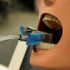
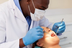
 A dental student works on a plastinated specimen. All images courtesy of UNLV Photo Services.
"You can move the specimens to get a better understanding of how one structure is deeper but also more lateral to another structure," said Dr. Joshua Polanski, an assistant professor in residence of biomedical sciences at the dental school. "That effect can be very hard to replicate with 3D-rendered images."
Some schools have started using plastinates instead of embalmed human cadavers, which are preserved using formaldehyde, for several reasons. Plastinated specimens are odorless, durable, and can last for decades. Though cadavers maintain more natural-feeling tissue texture, they can only be used for one semester.
In 2005, the New York University (NYU) College of Dentistry became the first dental school in the U.S. to use plastinated specimens for anatomy courses. Dr. Gunther von Hagens, a German anatomist who has been a visiting professor at the dental school since 2004, invented the technique of plastination in 1977.
NYU's specimens were produced at the von Hagens plastination laboratories in Germany and were derived from a body donation program that was established about 20 years ago. It has since registered about 19,000 donors worldwide via the Institute for Plastination in Germany. The program has reached its capacity and has stopped taking donations indefinitely.
Plastination gained traction when an exhibit of the technology was held in Tokyo in 1995. In 2016, the University of Texas Health Science Center at Houston School of Dentistry acquired plastinated specimens. Several universities and medical schools in the U.S. currently also use plastinates.
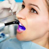
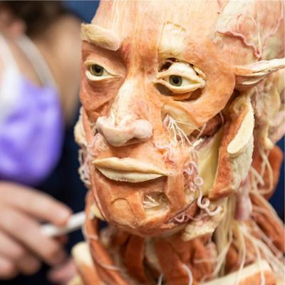
 Dental students work together with plastinated specimens.
UNLV acquired 60 plastinates and started using them to teach first-year dental students head and neck gross anatomy in the fall of 2020. They are also used in the school's neuroanatomy courses for first-year dental students and in a clinically oriented anatomy course for second-year dental students. Approximately 170 students will use the specimens per academic year, Polanski said.
"The plastinated specimens foster tactile experiences that lead to long-term retention of the curriculum," he said.
Tangible specimens also offer an edge over virtual anatomy learning tools. 3D virtual anatomy can be an effective supplement to using plastinated specimens or cadavers, but it is not a substitute, according to Polanski. A 3D replication is not of the same quality as tissue, and it fails to give students opportunities to mobilize joints, muscles, or organs, he added.
"Cadaveric learning demonstrates the durability versus the fragility of different tissues and offers the variation and pathology that virtual anatomy can never provide," he continued. "Each living patient is different, but most virtual anatomy app patients are exactly the same."