In this case, a 47-year-old female patient presented with congenitally missing upper laterals and microdontia. She was previously restored with bridges on teeth #6 through #8 and teeth #9 through #11, which were now fractured, stained, and exhibited leakage at the margins.
Clinical objective
The patient’s goal was to achieve functional and aesthetic improvement of her smile. Cone-beam computed tomography imaging determined that implants were not a viable option at sites #7 and #10. An iTero scan was performed to consider clear aligner therapy with Invisalign.
Upon considering possible treatment options, it was determined that the best union of aesthetic and conservative treatment would be to approach this case in three phases.
Phase 1: Orthodontic treatment would be used to reduce the thickness of bridge abutments, align the lower anterior spacing, intrude the anterior teeth to minimize reduction, expand the arches, and improve posterior occlusion. Clear aligner therapy took seven months to complete.
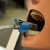
 An intraoral image taken after seven months of Invisalign treatment.
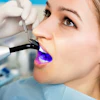
Phase 2: Whitening would be employed to attain the desired tooth shade. The patient chose to whiten her teeth with Opalescence gel and custom whitening trays.
Phase 3: New bridges for teeth #6-8 and #9-11 would be designed, fabricated, and seated. Additionally, there would be three-quarter crowns on teeth #23-26.
Technique
First appointment
Satisfied with the results of the whitening, the patient returned to the office to have her teeth prepped and provisionals placed. The teeth were minimally prepared, and digital impressions were taken with CEREC Primescan (Dentsply Sirona). In addition, pontic sites were surgically prepared on the day of preparation, and ovate pontics were designed using inLab Software.
 Following minimally invasive preparations, the patient was then fitted with 3D-printed provisionals.
Upon completion of all preparations, provisional restorations were 3D-printed using Flexcera Resin and the Envision One 3D printer. The provisionals were seated using temporary cement, and the patient was scheduled to return two weeks later for placement of the final restorations.
During the time between the first and second appointments, final restorations were fabricated and finalized in the dental office. The restorations were milled from Katana Zirconia One (Kuraray Noritake) CAD/CAM blocks in shade B1 on the CEREC Primemill. Post-mill and post-sinter contouring was completed, and the restorations were characterized with Cerabien ZR FC Paste Stains (Kuraray Noritake).
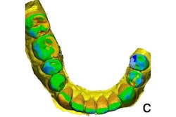
 The Katana Zirconia One bridge (teeth # 6-8 and #9-11) shown on a model after contouring/preglaze.
Second appointment
The patient returned to the office for a try-in and seating of the final restorations. The provisionals were removed and the preparations were cleaned of residual temporary cement. The final restorations were tried in and fit perfectly, with no adjustments necessary. The restorations were then bonded using Panavia SA Cement Universal (Kuraray Noritake), a self-adhesive cement.
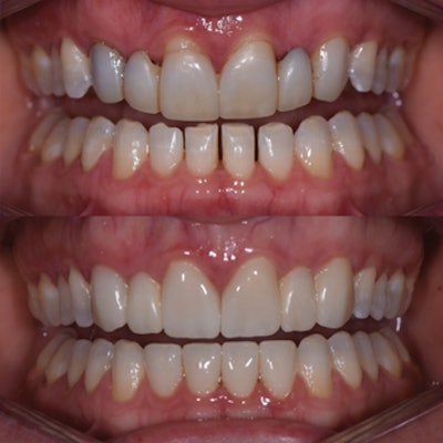
 Preop (top) and postop (bottom) of the patient. All images courtesy of Dr. Jenny Apekian.
Dr. Jenny Apekian is the founder and owner of Midtown Dental located in Sacramento, CA. She is a strong proponent of using the latest technologies in dentistry to improve the patient experience and has designed and built one of the most technologically advanced dental offices utilizing CAD/CAM technologies, 3D imaging, state-of-the-art infection control systems, as well as full digital integration.
The comments and observations expressed herein do not necessarily reflect the opinions of DrBicuspid.com, nor should they be construed as an endorsement or admonishment of any particular idea, vendor, or organization.