Ultrasound imaging has a promising future as a hard- and soft-tissue diagnostic tool in all dental specialties, according to a comprehensive literature review in the current Oral Surgery, Oral Medicine, Oral Pathology, Oral Radiology (June 2013, Vol. 115:6, pp. 819-832).
"Ultrasonography may provide a significant benefit to patients by allowing early detection of tooth lesions and defects, measurement of mucosa and gingival thickness, dental implant locations, and dental scanning," the study authors wrote.
For this review the international team of researchers conducted a Medline search from October 2011 to May 2012 using the terms "ultrasound," "ultrasonography," "image," and "dentistry" alone and in combination with one or more of the following terms:
- Hard tissue
- Soft tissue
- Dental implant
- Tooth
- Enamel
- Dentin
- Caries
- Fracture
- Cracks
- Periapical
- Lesion
- Gingiva
- Temporomandibular disorder (TMD)
- Muscle thickness
- Dental scanning
- Acoustic properties
From 4,125 articles identified by this search, the researchers ultimately selected 58 articles that met their criteria. Here are some of their findings:
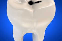
Caries detection: Of the studies that met their criteria, the researchers identified four studies that specifically looked at the use of ultrasound for caries detection.
For example, a 2003 study compared traditional bite-wing radiographs with ultrasound for diagnosing approximal caries and found that while ultrasound exhibited a higher sensitivity, it also yielded lower specificity (Oral Surg Oral Med Oral Pathol Oral Radiol Endod, May 2003, Vol. 95:5, pp. 626-631). The authors suggested that improving the ultrasound signal-processing algorithm might reduce the number of false positives and improve specificity.
Five years later, a team of U.S. and Turkish researchers compared ultrasonography to the Diagnodent (KaVo) and found that both methods demonstrated high repeatability and accuracy (Oral Surg Oral Med Oral Pathol Oral Radiol Endod, November 2008, Vol. 106:5, pp. 729-735). In addition, "all ultrasound measurements were accurate, reliable, and positively and significantly correlated between examiners," they wrote.
Soft-tissue lesions: Noting that "most dentists are unaware of the utility of ultrasound in the diagnosis of many types of oral diseases," which may be "disadvantageous to patients," the authors of the current study found five studies that assessed ultrasound's ability to diagnose these lesions.
For example, Japanese researchers in 2010 demonstrated the clinical applications of ultrasound in soft-tissue lesions, guided fine-needle aspiration, measuring tongue cancer thickness, and diagnosing metastasis to cervical lymph nodes (International Journal of Dentistry, 2010, doi:10.1155/2010/639382). "The Doppler mode ... was reported to be a useful modality in the differential diagnosis between normal and metastatic lymph nodes in patients with oral squamous cell carcinoma (OSCC)," the study authors concluded.
More recently, another team of Japanese researchers found that ultrasonography may be very useful for diagnosing postoperative stitch abscesses and thus help improve the quality of life of OSCC patients (Oral Oncology, March 2011, Vol. 47:3, pp. 163-169).
And just last year, investigators from M.M. College of Dental Sciences and Research conducted a 45-patient clinical study comparing ultrasonography with clinical diagnosis, radiography, and histopathology in diagnosing maxillofacial swellings (European Journal of Radiology, August 2012, Vol. 81:8, pp. 1821-1827). They found the diagnostic accuracy of ultrasound to be 92.3% in diagnosing cystic lesions, 87.5% in benign tumors, 81.8% in malignant tumors, 100% in lymphadenopathies, and 90% in space infections and abscesses. In addition, "significant results were obtained when ultrasonography was compared with clinical (0.895) and radiographic diagnosis (0.889)," the authors reported.
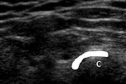
Periapical lesions: Because clinical examination and radiographs alone are not able to differentiate between cystic and noncystic periapical lesions, ultrasound is among a number of newer imaging techniques being studied for this application.
For example, a 2006 study compared ultrasound and conventional and digital radiography in diagnosing periapical lesions in 15 patients and concluded that ultrasound provided accurate information on the pathological nature of the lesions (Dentomaxillofacial Radiology, September 2006, Vol. 35:5, pp. 326-333). "The results showed that where sufficient buccal cortical bone had been resorbed, ultrasound imaging was straightforward but underestimated the size of the lesions" when compared with the other techniques, the authors reported.
Another study compared computed tomography scans with ultrasound imaging using power Doppler flowmetry (Journal of Endodontics, November 2008, Vol. 34:11, pp. 1312-1315). In all 12 cases included in the study, the researchers found that the diagnosis with computed tomography and ultrasound coincided with the histopathological diagnosis of the lesion. However, the authors noted, ultrasound is still considered a supporting technique and may not substitute histopathology at this point.
Similarly, a 2010 study compared the effectiveness of ultrasound, color Doppler imaging, and conventional radiography in monitoring postoperative healing of periapical lesions of endodontic origin and found that ultrasound with color Doppler was effective (Journal of Oral Science, September 2010, Vol. 52:3, pp. 411-416).
"This study showed that at six months, ultrasound and color Doppler imaging were significantly better than conventional radiography in detecting changes in the healing of hard tissue at the surgical site," the authors reported.
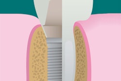
Periodontal bony disorders: In 2010, researchers from West Virginia University and Cairo University investigated the feasibility of using a custom-designed ultrasound imaging system to reconstruct 3D surface images of periodontal defects (Annals of Biomedical Engineering, November 2010, Vol. 38:11, pp. 3409-3422). Using this system, they were able to identify and describe in 3D all anatomical landmarks, while 2D radiographic images yielded poor contrast, the study authors noted.
"There is great potential for using high-resolution ultrasound as a noninvasive, nonionizing imaging technique for the early diagnosis of the more severe form of periodontal disease," they wrote.
And a literature review in the Journal of Periodontology found that ultrasound imaging is fast, noninvasive, and "is able to visualize periodontal and oral tissues in vivo or ex vivo without the need for complicated processing, fixing, or staining" (February 2010, Vol. 81:2, pp. 186-198).
Advantages of ultrasound
"The findings of this study present some of the advantages of ultrasound in comparison to traditional modalities, such as nonionizing radiation, noninvasive method, painless, accuracy, visualization of hard and soft tissue, and good acceptance by patients," the authors of the new study wrote. "Nevertheless, most dentists are still unaware of the full utility of this technology."
Further research should focus on achieving better quality image resolution for dental applications, they concluded.