Cone-beam computed tomography (CBCT) imaging helped clinicians use a novel surgical technique to remove a rare nasally impacted extra tooth in a 13-year-old boy. The case report was published on April 4 in Advances in Oral and Maxillofacial Surgery.
The teen’s nasally impacted asymptomatic mesiodens was surgically extracted successfully using a maxillary transnasal subperiosteal approach via a modified LeFort I vestibular incision. CBCT scans allowed for accurate 3D localization of the impacted tooth, allowing clinicians to devise better ways they can access challenging areas surgically, the authors wrote.
“This novel technique, which requires surgical experience and expertise, is recommended due to optimal surgical access, resulting in reduced surgical time and less bone removal with a decreased risk of damage to the adjacent structures,” wrote the authors, led by Dr. Peter Tsakiris of the Prince of Wales Hospital in Sydney.
A 13-year-old boy with a nasally impacted asymptomatic mesiodens
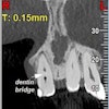
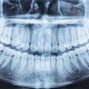
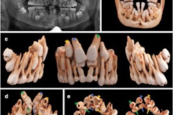
A healthy 13-year-old boy was consulted for preorthodontic surgical removal of an asymptomatic mesiodens in the nasal area. A CBCT scan and orthopantomogram showed that the extra tooth was located submucosally and breaching the nasal floor. Additionally, it was posterior to the boy’s upper right central incisor, the authors wrote.
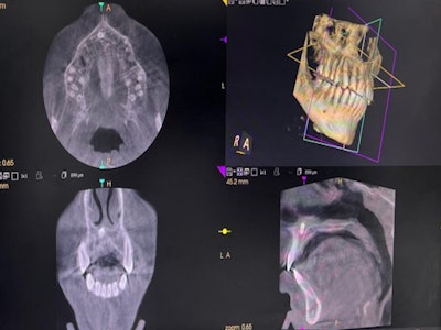
 A CBCT scan shows the teen’s nasally impacted tooth.Images courtesy of Tsakiris et al. Licensed by CC BY 4.0.
After the patient and his parents agreed to have the tooth extracted using the maxillary transnasal vestibular technique, the surgery was performed under general anesthesia with oro-endotracheal intubation.
“Through a LeFort I modified vestibular maxillary incision from the canine to the canine, subperiosteal dissection of the anterior maxilla and nasal floor was done, gaining access to the anterior aspect of the nasal floor,” the authors wrote.
The bone partially covering the mesiodens was removed, and the tooth was extracted. The patient recovered without complications, they wrote.
An alternative surgical strategy for an uncommon problem
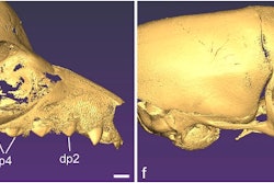
 The mesiodens following removal via a maxillary vestibular transnasal approach.
An upper jaw mesiodens is a common developmental anomaly, but it is rarely found inside the nasal floor. When an extra tooth is located on the nasal floor, it is often removed via palatal transalveolar or intraoral buccal approaches.
In 2011, a study described removing a nasally impacted mesiodens using a modified maxillary vestibular approach with subperiosteal intranasal dissection. Since then, this procedure has not been studied extensively or applied, according to the report.
Relevant skills in LeFort I osteotomy are necessary to handle the diagnostic, technical, and surgical requirements for this procedure, but the new approach offers advantages over the transalveolar approach, the authors wrote.
“[A modified maxillary vestibular approach offers] optimal visibility of the surgical field, time efficiency, decrease[d] risk of damage to the upper incisor roots and nasopalatine neurovascular bundle, resulting in minimal bone loss and elimination of the need to place a palatal splint post-operatively,” Tsakiris et al concluded.