Using new, modified dental imaging reconstruction parameters, cinematic rendering was implemented to visualize supernumerary and ectopic teeth separated from surrounding bone. The technical report was published on April 4 in Dentomaxillofacial Radiology.
Cinematic rendering, an alternative to volume rendering for 3D computed tomography (CT) scans, helps clinicians gain an improved understanding of a patient’s anatomical structures, allowing for better treatment planning. It is believed to be the first time this technique has been used to visualize teeth, since the software has yet to provide customized reconstruction parameters for dental imaging, the authors wrote.
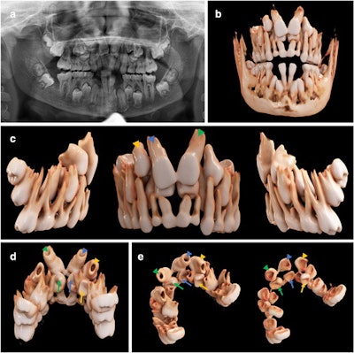
 An 11-year-old girl with a horizontally impacted canine (white arrows) and a persisting deciduous canine (white arrowheads). (a) An x-ray of the girl’s mouth. (b) Semitransparent reconstruction parameters are used to visualize bone, teeth, and different dental tissues. (c and d) The teeth and bone tissue with a soft kernel show a photorealistic visualization in both a frontal and lateral view.
“When employing our new, modified reconstruction parameters, its application presents a fast approach to obtain realistic visualizations of both dental and osseous structures,” wrote the authors, led by Dr. Ines Willershausen of the department of orthodontics and orofacial orthopedics at Friedrich-Alexander-Erlangen-Nurnberg University in Germany.
Recently, cinematic rendering, a technology that uses data derived from CT and cone-beam CT images, has been used as an alternative approach for visualizing volumetric medical imaging data. It is a novel visualization technique using physically based volume rendering to create photorealistic images from Digital Imaging and Communications in Medicine (DICOM) data.
 The postprocessing pipeline of volumetric data for 3D visualization by means of cinematic rendering.
Cinematic rendering builds on novel, physically based light simulation algorithms in which billions of virtual light rays are simulated every second in a physically accurate manner. The technology offers a more realistic appearance than images created via conventional volume rendering. Typically, the conventional method only uses a single directional light source, making images look less lifelike, the authors wrote.
Since its introduction, cinematic rendering has found applications in medical clinical research and anatomical education, but it has not been tailored to dentistry. Currently, there is only limited research on this technology in dentistry. Mainly, cinematic rendering has focused on fractures and carcinoma as maxillofacial indications, the authors of the report wrote.
In the report, researchers used cinematic rendering for the volumetric image visualization of midface CT datasets. Predefined reconstruction parameters were specifically modified to visualize complex orthodontic cases in children with ectopic, impacted, and supernumerary teeth. Using the masking and windowing functionality of Siemens Healthineers’ Cinematic Anatomy application, upper and lower jaw and respective dentitions were easily segmented, creating natural-appearing images for realistic representations of anatomy, the authors wrote.
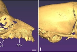
 An 11-year-old boy with a mesiodens and a supplementary tooth (white arrows) within the hard palate. (a) An x-ray of the boy’s mouth. (b) A display of the semitransparent reconstruction parameters. (c and d) The bone reconstruction parameters with a soft kernel can lead to artifacts in regions with teeth near each other (yellow circle in d, e, and f.) The same situation is shown with a hard kernel, allowing better differentiation between the roots of the mesiodens and of tooth #21 (yellow circle in f).
Also, the current reconstruction time of up to 24 hours is not reasonable for routine clinical applications. However, as the software undergoes updates, reconstruction time is expected to significantly decrease, they wrote.
“(Nevertheless,) the 3D spatial relationship of the teeth, as well as their structural relationship with the antagonizing dentition, could immediately be investigated and highlighted by separate, interactive 3D visualization after segmentation through windowing,” Willershausen and colleagues wrote.