Imaging helped lead to the diagnosis and treatment of a 2-year-old girl who developed the life-threatening condition orbital cellulitis due to a dental abscess. The details were published on April 9 in the Cureus Journal of Medical Science.
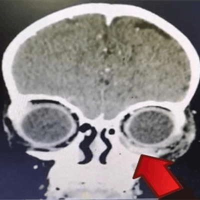
A dental exam revealed the child had multiple caries, gum swelling, and loose baby teeth, and a contrast-enhanced computed tomography (CT) scan showed a left periosteal abscess collection extended to her orbit. A dental abscess can cause the serious bacterial infection orbital cellulitis, and this case highlights the importance of early tooth care in children, the authors wrote.
"The intraoperative results showed the presence of a left upper gum abscess, which was possibly the primary source of infection," wrote the group, led by Dr. Huwaina Satar of the department of ophthalmology and visual science at University of Science Malaysia School of Medical Sciences.
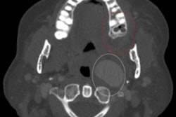
 The arrow indicates the periosteal abscess collection extending into the inferomedial aspect of the left orbit. All images courtesy of Satar et al. Licensed under CC BY-NC 4.0.
Life-threatening but treatable
Staphylococcus, Streptococcus species, and Haemophilus influenzae are the most common types of bacteria responsible for pediatric orbital cellulitis, and sinusitis and trauma are the most common risk factors for it. However, the condition is also a rare but serious complication of a dental infection. If orbital cellulitis is not treated, it can spread to the lymph nodes and bloodstream and quickly become deadly.
The child was brought to the hospital after having a low-grade fever and progressive left periorbital swelling for three days. The swelling first appeared on the left upper perioral region and extended upward to the cheek and lower lid on the left side. She had no history of trauma, and her eye was not red or watery, the authors wrote.
 The progression of the swelling, which worsened on the third day after admission, of the girl's left eye.
The girl was active and had stable vital signs. An eye exam revealed left eyelid redness and a firm left periorbital swelling. All eye exams were normal, and her eye swab and blood cultures were negative.
The patient was admitted to the hospital, diagnosed with preseptal cellulitis, and given intravenous antibiotics. Doctors also prescribed an antibiotic cream for her eye.
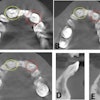
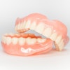
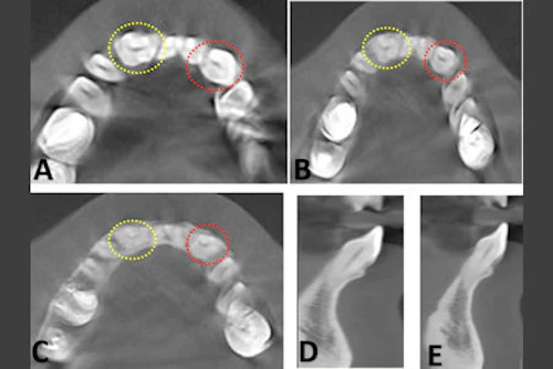
 A dental exam showed multiple dental caries and upper gum abscess (arrow).
On her third day in the hospital while still be treating with antibiotics, the eye swelling increased. A dental exam showed multiple dental caries, upper gum swelling and an abscess, and two loose baby teeth, according to the authors.
Contrast-enhanced CT of the orbit, paranasal area, and brain showed a left periosteal abscess collection that extended into the left orbit and maxillary and ethmoidal sinuses. A dental exam and tooth extraction were completed while the girl was under general anesthesia, and the dentist discovered a left upper gum abscess. A culture was taken, and no organism was isolated.
 The orange arrow indicates the gum swelling and the green arrow indicates the abscess collection.
The primary source of the infection was traced back to the area near her two loose teeth, which showed early swelling at the upper perioral region. The infection then moved into her sinuses and eye, the authors explained.
The girl completed one week of intravenous antibiotics, her swelling decreased, and she was discharged.
The case shows that imaging is pivotal to diagnosing and treating orbital inflammation, especially when the condition worsens. Additionally, the case is important because it shines a light on a rare complication of a dental infection, the authors wrote.
"Awareness of the possible spread of odontogenic infection to the orbit is extremely important, so vigorous treatment may start as early as possible," Satar and colleagues wrote.