Research presented by the University of Maryland Dental School at this week's Microscopy Society of America annual meeting in Richmond, VA, showed cylindrical objects extending out of the natural pores or tubules of teeth, according to the university.
 |
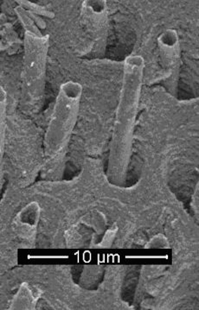
| Scanning electron microscope image of wormlike structures 'growing' from dental tubules deep inside a molar. Image courtesy of University of Maryland, Baltimore. |
Inside a human tooth, more than 50,000 such tubules per square millimeter act as channels running from the nerve up through the tooth. They are associated with transporting hot or cold sensitivity to the tooth nerve.
The aim of the Maryland study was to investigate the structures with scanning electron imagery and different specimen preparation techniques, according to the university.
While the tubules ranged from 2.6 to 3.5 micrometers (µm) in diameter, the structures were as long as 9 µm, extending out of the tubule opening. While most of the structures appeared to be hollow, a number of them appeared to be solid.
For years, scientists have debated the exact nature of the worm-like structures, which were photographed in clear detail by Ru-Ching Hsia, director of the electron microscope core facility at the school.
Dentists have differing theories on these structures.
"Most say, 'I have no idea.' Others say they are made of bacteria, or minerals, or hyphal branches of yeast cells (Candida albicans), which have infected the tooth structure, or perhaps they are a cellular process of the dentinal tubules," said co-presenter Gary Hack, D.D.S., an associate professor at the dental school. For the sake of humoring his students, Dr. Hack said, "I call them tooth worms and I'm sticking to it."
Copyright © 2009 DrBicuspid.com