Endoscopic narrow-band imaging (NBI) is more effective than broadband white light (BWL) imaging in detecting high-grade dysplasia and carcinomatous lesions in oral leukoplakia, according to a new study in the International Journal of Oral & Maxillofacial Surgery (April 11, 2013).
For the study, a team of researchers from Taiwan investigated the clinical efficacy of using BWL to observe morphologic appearance, NBI to observe intraepithelial microvasculature, and both to detection high-grade dysplasia and carcinoma in oral leukoplakia. They wanted to see if, when used to assess oral leukoplakia, NBI can provide more information than the traditional classification under BWL.
"Narrow-band imaging is an innovative optical technology that enhances the contours and patterns of vessels, or intraepithelial microvasculature, in the surface of mucosa by employing the characteristics of the light spectrum," the study authors wrote.
They reviewed the records of 317 patients at Chang Gun Memorial Hospital who were newly diagnosed with oral leukoplakia between April 2009 and August 2011, were examined using flexible endoscopy with BWL and NBI, and underwent biopsies or surgical excision for the leukoplakia.
The optical exams were conducted using NBI endoscopes, light sources, and a central video system from Olympus Medical Systems. A button on the control section of the video-endoscope allowed for switching between the BWL and NBI views.
"The examinations were first conducted using BWL illumination in a wide view to observe the entire lesions and its surrounding mucosa," the researchers explained. "The same procedure was performed under NBI illumination for detailed analysis of the appearance of the intraepithelial microvasculature and screening of random sites of normal mucosa."
The resulting images were then analyzed and compared with the histopathologic finding for each patient. The clinical morphologic appearance under BWL was analyzed first, including homogeneous and nonhomogeneous leukoplakia. The latter was categorized as the verrucous, nodular, or speckled type. Imaging analysis of the NBI features (microvascular organization and intraepithelial papillary capillary loop) was conducted based on the intraepithelial microvasculature patterns of the oral mucosa.
Sensitivity, specificity, false positives
Each patient's chart records were reviewed, including their demographic data, lesion location, homogeneous/nonhomogeneous leukoplakia, vascular architecture, and histopathology. Using the histopathologic findings, the researchers then determined any correlation between pathology and imaging by BWL and NBI by assessing the sensitivity, specificity, positive and negative predictive values, accuracy, and false-positive and false-negative percentage in detecting high-grade dysplasia, carcinoma in situ, and invasive carcinoma (HGD/Tis/CA). They also compared traditional classification of leukoplakia based on morphological appearance using BWL and pattern of intraepithelial microvasculature using NBI.
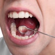
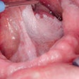
"Observing oral leukoplakia with the naked eye is not difficult, but the reddish component of nonhomogeneous leukoplakia, especially the speckled type, may not be prominent and may be neglected or mistaken for homogeneous leukoplakia," the researchers noted.
Using an endoscope with improved brightness, light distribution, high-resolution images with better color reproduction capability, and larger display size, "white plaque lesions can be examined at close range, in detail, to reduce this inaccuracy," they added.
Among the 317 patients analyzed in this study, the researchers found that using NBI classification based on the intraepithelial microvascular patterns was significantly better than traditional classification in detecting HGD/Tis/CA (p < 0.001). Although the sensitivity and negative predictive value of traditional classification were better than NBI (96.30% and 97.85% versus 87.04% and 97.23%), "the specificity and diagnostic accuracy of the former are much poorer" (60.08% and 66.25% versus 93.54% and 92.43%). In addition, the false-positive rate of traditional classification in detecting HGD/Tis/CA of oral leukoplakia is higher than NBI classification (39.92% versus 6.46%), they noted.
Comparison between the traditional BWL classification and NBI classification for detecting HGD/Tis/CA of oral leukoplakia was significant (p < 0.001), while comparison between the traditional BWL classification and combined BWL and NBI classification was not (p = 0.564). However, comparison between the NBI classification and combine BWL and NBI classification was significant (p < 0.001).
"The NBI classification based on intraepithelial microvascular patterns is significantly better at detecting HGD/Tis/CA lesions in oral leukoplakia than the traditional BWL classification based on the morphological appearance," the study authors wrote. "As endoscopy with NBI can provide useful data regarding dysplastic or carcinomatous changes, it is a promising tool in detecting HGD/Tis/CA of oral leukoplakia."
This is not the first study the research team has published demonstrating the efficacy of endoscopy and NBI to evaluate oral leukoplakia. A study published last year in Head & Neck (July 2012, Vol. 34:7, pp. 1015-1022) found that the correlation between intraepithelium papillary capillary loop classification and stepwise increased severity of pathology was significantly better using NBI than BWL images (p < 0.001).
"Flexible endoscopy can enhance detailed inspection of oral cavity mucosa and can be a powerful tool for examining oral leukoplakia," the authors concluded.

