The chances of encountering nonendodontic lesions in periradicular tissues are nearly 8%, and up to 3% of biopsies conducted on teeth with pulp necrosis were confirmed to not be of an endodontic origin, according to a study published in the Journal of Endodontics.
Since these findings were established after comprehensive clinical exams, histological investigations are needed to obtain a definitive diagnosis, according to an 18-year retrospective study. It is believed to be the first study to identify nonedodontic lesions obtained from endodontic microsurgeries, the authors wrote.
“Pathologic findings of the periradicular tissues are not always from endodontic origin,” wrote the authors, led by Dr. Fabricio Teixeira, PhD, of the department of endodontics at the University of Iowa College of Dentistry in Iowa City (J Endod, August 2, 2023).
Looks can be deceiving
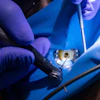
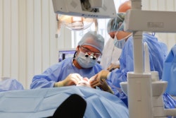
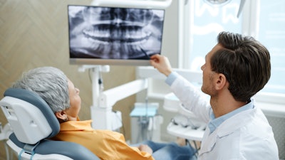
Infected root canal systems are constantly linked to imaging findings in patients’ periradicular areas. Clinically and on imaging, nonendodontic lesions can appear similar to endodontic lesions, creating challenges in differentiating between the two types. Not identifying the correct lesion can result in unsuccessful endodontic surgeries. To better plan patients’ treatments and to avoid misdiagnoses, clinicians may have to consider additional diagnostic strategies like histopathology tests prior to microsurgeries, according to the study.
To improve the accuracy of findings that can contribute to a more informed understanding of periapical lesions that may mimic pathologies with endodontic origins, the authors reviewed biopsies submitted to the University of Iowa’s College of Dentistry and dental clinics between 2003 and 2021 were reviewed. Histopathologic diagnosis and gross description were also available for exploration, they wrote.
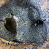
Of the 72,055 pathology reports reviewed, 10,031 lesions (13.9%) were considered intraosseous lesions in the periradicular area. Among those lesions, 796 (7.9%) were of nonendodontic origin, the authors wrote. Additionally, 7,153 were documented as nonvital, and 169 (2.4%) of these nonvital teeth were diagnosed with nonendodontic origins, the authors wrote.
Overall, the most common nonendodontic diagnosis was odontogenic keratocyst followed by nasopalatine duct cyst and benign fibro-osseous lesion, respectively. As for malignancies, the most frequent found at the periraridcular area were hematopoietic malignancies followed by squamous cell carcinomas, plasma cell neoplasms, metastatic carcinomas, adenocarcinomas, Langerhans cell histiocytosis, and malignant neoplasms not otherwise specified, the authors wrote.
A limitation of the research was that four times more biopsies were collected from the upper jaw than the lower jaw in the study's samples. Since most nonendodontic lesions are found in the mandible, the frequency of these lesions obtained from surgery may be underestimated, the authors wrote.
The importance of lab testing
Multiple types of nonendodontic lesions may look like endodontic lesions on imaging, since the incidence of nonendodotic lesions in the periradicular area was about 8%. Though an editorial published in November 1998 in Oral Surgery, Oral Medicine, Oral Pathology, Oral Radiology, and Endodontics suggested that biopsies may be avoided if clinicians conduct extensive clinical exams and carefully evaluate patients’ medical histories, the findings of the current study may indicate otherwise, they wrote.
“Therefore, clinicians must perform a thorough clinical examination and obtain the definitive diagnosis before initiating treatment to reduce the potential for misdiagnosis and subsequent errors in treatment planning,” Teixeira et al concluded.