A rare dental developmental abnormality, known as a protostylid and characterized as an extra cusp, caused a woman’s apical periodontitis, according to a case report published on August 28 in BMC Oral Health.
Cone-beam computed tomography (CBCT) scans allowed clinicians to visualize the supernumerary cusp with an intact root canal, which was fused with the mesiobuccal canal in the middle of the root. The woman underwent endodontic treatment and was clinically asymptomatic after one year, the authors wrote.
“Protostylid is rare to be reported, and the fusion of the mesial buccal cusp with a protostylid associated with root canal treatment has not yet been reported,” wrote co-author Dr. Ye Tao of the hospital of stomatology at Sun Yat-Sen University in Guangzhou, China.
A 53-year-old woman experiencing pain during chewing
The woman, who had no significant contributing medical history, presented with a primary complaint of three months of pain in her upper left posterior teeth when chewing.
During an oral examination, an eroded cusplike structure was found on tooth #26’s buccal surface. The cusp extended from the cervical edge of the tooth toward the mesiobuccal cusp. The tooth did not respond to an electric pulp and cold test and was preliminarily diagnosed with pulp necrosis, the authors wrote.
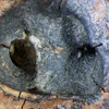
 (a, b) Preoperative image showing occlusal and buccal planes of the protostylid in tooth #26. The red open circle indicates the protostylid. (c) A panoramic view of permanent teeth with normal anatomy. Images courtesy of Tao et al.
Dental x-rays revealed an extensive periapical radiolucency around tooth #26, but it was unclear whether the protostylid had its own root canal. However, a CBCT image revealed the complex anatomy of the tooth. The pulp chamber of the protostylid was independent in the anterior third of the root and fused with the mesiobuccal root canal in the apical half of the root. Therefore, the patient’s tooth was diagnosed with apical periodontitis, they wrote.
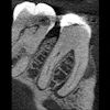
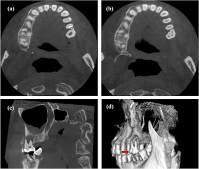
 CBCT images (a, b) of the cross section of the plane of the protostylid in tooth #26. (c) The vertebral plane of the protostylid in tooth #26. (d) A 3D reconstruction of the protostylid in tooth #26. Red solid arrows indicate the protostylid.
Since the tooth had extensive cortical bone destruction of cortical bone, endodontic treatment was planned. When the clinician began working on the tooth, it was discovered that the tooth had five root canals. There was a mesiobuccal canal, a second mesiobuccal canal, the distobuccal canal, the palatal canal, and the root canal in the protostylid, according to the report.
After treatment was completed successfully, the woman was scheduled for follow-up visits. At the one-year follow-up visit, the tooth remained free of symptoms, and imaging showed the tooth had a healing apical area.
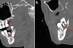
 Immediate postoperative and follow-up imaging. (a) The filled root canal under an operating microscope. (b) A 3D reconstruction of filled tooth #26. (c) A periapical radiograph taken after treatment. (d) A radiograph taken at the six-month follow-up. (e) A radiograph taken one year after the root canal.
An extra cusp requiring special consideration
Usually, a supernumerary cusp is an accessory cusp on the buccal or lingual surface of a normal crown. Typically, a protostylid appears as a pit and distal bending of the buccal groove or surface abnormality. However, in this case, the protostylid was located on the mesial half of the buccal surface on the woman’s first molar.
Though the origin of the protostylid is unknown, it has been theorized that they originate from the growing center of an enamel knot that eventually becomes the location of the cuspid vertices during the morphogenetic phase of tooth development.
Since it is difficult to differentiate between a protostylid and a fused tooth and a tooth with concrescence, its diagnosis should be determined carefully. Additional imaging like CBCT is useful in making the distinction, they wrote.
“In conclusion, the occurrence of protostylid is a relatively rare phenomenon,” Tao and colleague wrote.