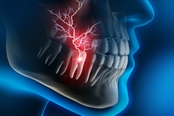
Recently, researchers used magnetic resonance imaging (MRI) to measure brain activity in patients with bruxism and temporomandibular disorder (TMD)-related pain, according to research published in the Journal of Oral & Facial Pain and Headache.
A team led by Dr. Theo Kluskens of Maastricht University in the Netherlands reported that brain activity in patients with bruxism and TMD-related pain was tied to pain processing more so than motoric differences.
The researchers measured brain activity in patients with pain related to these disorders and compared such activity to controls by using functional MRI. They also wanted to learn whether changes in jaw clenching led to different pain reports or changes in neural activity in the motor and pain processing areas of the brain.
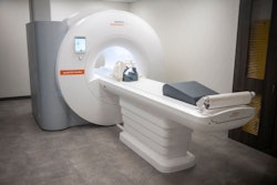
The team investigated brain activity in 40 patients, 21 of whom had bruxism and TMD-related pain, while the remaining 19 patients served as healthy controls. The patients performed a tooth-clenching task while lying inside a 3T MRI scanner. They were told to mildly or strongly clench their teeth for 12 seconds and to rate their clenching intensity and pain experience after each clenching period.
Patients reported significantly more pain during strong clenching compared to mild clenching. Furthermore, the researchers reported significant differences between TMD patients and control participants in brain network activity commonly connected to pain processing, which were also correlated with reported pain intensity (J Oral Facial Pain Headache, Spring 2023, Vol. 37:2, pp. 139-148).
The team, however, reported no evidence of differences in activity in motor-related areas between groups. It noted that this finding clashes with conclusions from previous studies.