Imaging aided in the retrieval of dentures from the bowel of a mentally competent 52-year-old woman who unknowingly swallowed them, according to a case report published online January 28 in the International Journal of Surgery Open.
A 3D reconstruction of a computed tomography (CT) scan allowed clinicians to identify that she had swallowed a sharp, hooked object, which guided their surgical planning. Also, this case highlights the fact that populations other than vulnerable ones are at risk of ingesting dentures, the authors wrote.
This case "raises awareness to the fact that the diagnostic is often elusive since even mentally capable patients might not notice or refer about ingestion of foreign bodies, especially dentures," wrote the group, led by Dr. David Salvador of the department of general surgery at Hospital Dr. José Maria Grande in Portalegre, Portugal.
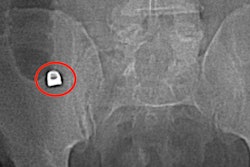
 CT scan shows a dense metallic object located in the small bowel in the right lateral quadrant with free fluid and localized fat thickening. All images courtesy of Salvador et al. Licensed under CC BY 4.0.
Foreign body ingestion is a common occurrence in the emergency department, but it often occurs in children, the elderly, or adults with cognitive delays or mental health conditions. To prevent serious complications, including intestinal perforation or obstruction, clinicians need to have thorough discussions with patients and use imaging to locate the foreign body and choose the right treatment plan, according to the authors.
In the current case, a 52-year-old woman went to the emergency room after 14 hours of gradual, generalized abdominal pain and nausea. She had no fever, vomiting, or other symptoms. She had a history of diabetes, high blood pressure, and high cholesterol.
Examination revealed tenderness in the woman's right iliac fossa, as well as inflammation of her peritoneum. Lab results showed an increased white blood cell count and C-reactive protein level. A chest x-ray showed no sign of pneumoperitoneum.
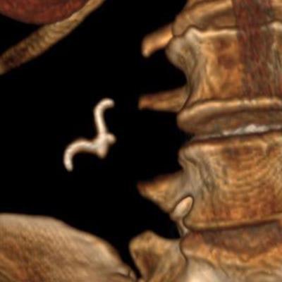
 3D CT reconstruction shows a hook-shaped object, which turned out to be the woman's dentures.
An abdominal and pelvic CT scan showed an endoluminal metallic object in the right lower quadrant, and clinicians asked the patient whether she had swallowed a fish bone or ingested any foreign objects, including dentures. She did not remember ingesting anything unusual, the authors wrote.
To identify the object and potential for perforation, they obtained a 3D CT reconstruction. The reconstruction showed the foreign object had a sharp hook that could perforate the bowel loop, so they opted to remove it surgically.
The surgeon immediately felt a hard object upon touching the patient's bowel loops. A closer look revealed an object impaled in the wall of the ileal bowel showing signs of impending perforation, approximately 20 cm from the ileocecal valve. The object, which was identified as a partial denture, was successfully retrieved.
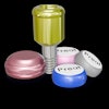
 The patient's surgically removed partial denture.
Fortunately, the woman's postoperative recovery was uneventful. She was discharged six days after surgery, the authors wrote.
Clinicians should ask patients with abdominal pain -- regardless of mental health status -- about possible foreign body ingestion and follow up with imaging to rule out other conditions, they concluded.
"When presented with a patient with unexplained abdominal pain, the ingestion of foreign object should be present in the differential diagnosis even in a mentally capable patient," the authors wrote.