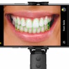
When you use an intraoral camera in your practice, do you use an orange filter and assess the red emissions from bacterial deposits as many practices do? A new study from Australia suggests that using violet or blue illumination combined with green compensating filters may be a better fit to judge mineral loss and make other clinical assessments.
Under ultraviolet, violet, and blue light, human enamel has a natural green fluorescence. Many studies use a broadband violet-blue light to assess any changes in green fluorescence, according to the study authors.
"The results of this study provide the groundwork for further exploration of imaging systems and devices that could be used in clinical settings," wrote authors Fardad Shakibaie, MDSc, PhD, and Laurence J. Walsh, DDSc, PhD, from the University of Queensland School of Dentistry (Australian Dental Journal, January 28, 2016).
Capturing images
Intraoral cameras are used for more than viewing and capturing images of tooth surfaces. They can also provide real-time education of patients during a consultation and enhance knowledge of the caries process, hopefully engaging patients more strongly in prevention and treatment.
“The results of this study provide the groundwork for further exploration of imaging systems and devices that could be used in clinical settings.”
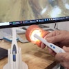
Intraoral cameras that have violet or blue light sources use orange filters, the authors noted. These are designed to assess green fluorescence of soft tissues, and the authors cited the VELscope (LED Medical Diagnostics) as one example. In this study, however, the researchers looked at the use of a "selective green filter stack when used with violet or blue light to assess green fluorescence of a tooth's structure."
"This filtration emphasizes the natural green fluorescence emissions from sound tooth structure and the lack of fluorescence from areas of dental caries," the authors wrote.
With the goal of comparing violet and visible blue LED light-elicited green fluorescence emissions from enamel and dentine in healthy or carious states, the researchers selected a total of 132 extracted human permanent teeth from a large repository of extracted teeth. The teeth were divided into eight groups. They also noted how green fluorescence was affected by the presence of saliva or blood on the surface of the teeth.
| Sample groups | |||
| Group | Number | Type of teeth | Storage material |
| A | 10 | Sound teeth | Diluted NaCIO |
| B | 10 | Sound teeth (teenage) | Diluted NaCIO |
| C | 20 | Dentine caries (dark color) | Diluted thymol |
| D | 20 | Dentine caries (not colored) | Diluted thymol |
| E | 20 | Sound root surface with plaque | Diluted saliva |
| F | 20 | Sound enamel with plaque | Diluted saliva |
| G | 6 | Tetracycline-stained teeth | Diluted thymol |
| H | 6 | Root-treated stained teeth | Diluted thymol |
Using violet and blue LED illumination (405- and 455-nm wavelengths) for excitation light of the tooth surfaces, the researchers photographed the teeth with microscopic digital photography through a custom-made filter stack of green compensating filters. The stack removed blue, violet, and red light, so that they could compare green fluorescence from sound and carious teeth, as well as teeth with intrinsic stains.
Sound enamel and root surfaces which were free of fluid registered a "strong green fluorescence" when excited by violet light or blue light, but regions of cavitated dental caries had a lower green fluorescence, the authors reported. The presence of saliva on the surface did not significantly change the fluorescence, but the presence of blood diluted in saliva did depress the green fluorescence.
Teeth with caries had green luminosity scores of less than 114 under violet excitation and less than 178.9 under blue excitation, compared with noncarious samples that consistently averaged scores of up to 250. The authors also noted that teeth with root fillings or tetracycline stains were not significantly different from sound teeth, but they showed a wider range of readings.
Saliva's influence
The authors did address what influence, if any, that ambient fluids covering the tooth's surface may have. "Saliva did not significantly affect the green fluorescence properties of dental samples compared to the fluid-free situation," they wrote. They noted, however, that issues arose with whole venous blood because of its strong absorption and high optical density for blue light.
Overall, the study showed the potential value of a selective green filter stack when used with violet or blue light to assess green fluorescence of tooth structure, the authors concluded. This filtration emphasizes the natural green fluorescence emissions from sound tooth structure and the lack of fluorescence from areas of dental caries.
"The present study shows that a simple approach using violet or blue LEDs and a stack of commercially available filters can be used with a digital camera to assess such light fluorescence properties," they wrote.