Panoramic x-rays may not be enough to get an accurate view of residual bone that is available for an implant when the maxillary sinus is tiny and alveolar resorption is significant, according to a brief case report that was published on August 22 in Head and Face Medicine.
In such instances, cone-beam computed tomography (CBCT) should be used to get the precise measurement of available residual bone to prevent an implant from perforating a patient’s nasal cavity, the authors wrote.
“Therefore, the residual bone between the lateral wall of the nasal cavity and the medial wall of the maxillary sinus should be reevaluated three-dimensionally to measure the exact bony shape and volume,” wrote the authors, led by Dr. Jo-Eun Kim of the Seoul National University School of Dentistry in Korea.
To get a better understanding of what factors may be associated with potential nasal cavity perforation by dental implants, the authors reviewed the cases of three patients. Through the review, they aimed to identify significant panoramic imaging characteristics of the nasal cavity and their relationship with the adjacent maxillary sinus.
A 51-year-old woman
The patient was referred to the hospital after an implant migrated to her superior cavity. An x-ray revealed that one implant that was intended for the premolar region had been displaced horizontally between the boundaries of the nasal cavity and maxillary sinus, appearing to have perforated the medial wall of the maxillary sinus. A CT image confirmed that the implant had migrated to the nasal cavity.
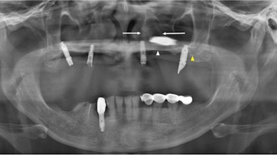
 The panoramic x-ray of a 51-year-old woman. The white arrow indicates the left hard palate line, while the yellow arrow indicates the antral floor. The implant fixture is displaced into the superior cavity, but the bone between the white arrows appears to have sufficient volume. All images courtesy of Kim et al. Licensed by CC BY 4.0.
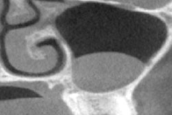
 The CT image of the first patient shows that the implant was displaced in the left nasal cavity but not in the maxillary sinus (a-c). The implant of the right premolar area, which appeared to be placed in a spot with insufficient bone between the nasal cavity and the maxillary sinus on an x-ray, appears to penetrate the nasal floor.
A 64-year-old man
The man was referred to the hospital for an oronasal fistula. The patient complained that his dental implants had failed three times.
An x-ray revealed an implant retained in the maxillary right canine area just lateral to the right nasal cavity. A CBCT scan showed obvious discontinuity of the inferolateral cortex of the nasal cavity and oronasal fistula. This was observed as a tissue defect on panoramic x-ray, according to the report.
 The panoramic x-ray of a 64-year-old man shows that perforation is suspected at the right maxillary sinus floor (white arrow). The white arrow indicates the left hard palate line, while the yellow arrow indicates the antral floor.
 The 64-year-old's CBCT images showed a discontinuity of the inferolateral cortex of the nasal cavity and oronasal fistula, which is observed as a tissue defect on the x-ray. (a) Moreover, the residual implants on both sides perforated the nasal cavity and are placed just medial to the maxillary sinus.
A 76-year-old woman
The woman had pain in the mandibular right posterior region and was diagnosed with osteomyelitis of the right posterior mandible based on clinical and imaging exams. CT images showed her dental implants had perforated the nasal floor of the maxillary left premolar.
However, panoramic x-rays revealed that the implants of the left maxillary premolar region were placed normally between the lateral wall of the nasal cavity and the faint medial wall of the maxillary sinus, the authors of the case study wrote.
 The x-ray and CT images of a 76-year-old woman. On the x-ray, white arrows indicate the hard palate line, while yellow arrows indicate the antral floor. Images of the implants in the left premolar and molar areas are superimposed above the hard palatal line. (b-c) The CT images revealed nasal floor perforation by an implant of the maxillary left premolar.
Common denominators
After reviewing the three cases, common features were discovered. First, the horizontal radiopaque line of the hard palate appeared to be inferior, or like that of the antral floor. Also, the bone between the nasal cavity’s lateral wall and the maxillary sinus’ medial wall was emphasized in a triangular shape, the authors wrote.
Why imaging matters
When considering implant placement in the arch of the anterior region of the upper jaw, the nasal cavity is a critical landmark. Precise assessment of the nasal cavity is required to avoid dental implants from perforating the area and causing complications, including inflammation and implant migration, the authors wrote.
Since panoramic x-rays are used because they are inexpensive, readily available and offer high-resolution images, they often are used to visualize the jaw for implant placement. However, due to the limitations of these two-dimensional images, it can be difficult to measure the ideal position and anatomical structure in the buccolingual direction, Kim and colleagues wrote.
"If three-dimensional structural features cannot be sufficiently determined from the acquired panoramic radiograph, three-dimensional imaging should be acquired accordingly,” they wrote.