Will Raman spectroscopy one day find its way into the dental operatory as an oral health assessment and caries detection tool?
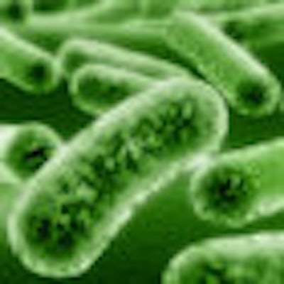
A team of University of Rochester scientists is using this highly sensitive microscopy technique to study two common dental plaque bacteria: Streptococcus sanguinis and mutans. The relative balance of the two may be an indicator of a patient's oral health and risk for tooth decay; S. sanguinis is associated with "healthy" plaque, while S. mutans is associated with tooth decay.
The researchers presented their findings October 26 at Frontiers in Optics 2010, the Optical Society of America's annual meeting in Rochester, NY. They also describe their research in the November/December 2010 Journal of Biomedical Optics (Vol. 15:6).
Why Raman?
Compared to microbiology techniques, Raman spectroscopy offers the potential to analyze samples of the bacteria in a simple, rapid, and quantitative manner, including the ability to study spatial distributions of bacterial species, living or dead, within samples, according to the researchers.
“Raman can be more specific than fluorescence.”
— Brooke Beier, University of Rochester
Institute of Optics
Since Raman spectroscopy is a scattering technique, specimens do not need to be fixed or sectioned. In addition, Raman spectra can be collected from a very small volume (< 1 µm in diameter); these spectra allow the identification of species present in that volume. And water does not generally interfere with Raman spectral analysis, making it suitable for examining proteins, cells, and organs.
For these reasons, Brooke Beier, a doctoral candidate at the University of Rochester Institute of Optics, and colleagues opted to use confocal Raman microspectroscopy rather than fluorescence or other analytical tools to study these two oral microbes.
"When it comes to looking at differences between species, Raman can be more specific than fluorescence if you're not using exogenous labels," she told DrBicuspid.com. "You can pick up on differences in a more natural form, and it is more sensitive to site changes in proteins."
To distinguish between biofilm samples of S. sanguinis and S. mutans, the researchers calibrated a species-identification model with spectra from pure biofilms and validated the model on a unique set of pure biofilms grown at a later date. They also examined the spatial resolution at which correct classifications can be performed in a multispecies environment.
"Examining the relative concentration balance between these two species may serve as an indicator of a patient's oral health and risk of tooth decay," they noted.
Species-specific distinctions
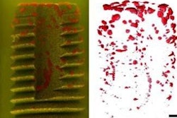
When they compared white-light transmission images of the biofilm -- in which it was not possible to distinguish between the two species based on appearance alone -- to the data obtained using the Raman microspectrometer, the researchers found that Raman spectroscopy's chemical specificity offers the ability to distinguish between the species.
Species-specific distinctions in Raman spectra are most likely a result of genomic differences between organisms, they noted.
"We're using Raman spectroscopy to study these oral bacterial biofilms, essentially observing how two species scatter light into shifted wavelengths in a unique way," Beier said. "We can then use these characteristic spectra to identify 'unknown' samples of these species."
This approach might also be useful in identifying the balance between these two bacteria so that practitioners could flag a patient as being at risk for oral disease, she added.
"If a patient has too much mutans, for example, we might be able to catch that earlier and counteract it," she said.
With the ability to identify biofilm samples by species, the researchers can now move on to study biofilms grown from a mixture of liquid cultures, where the two species may interact as they grow together.
"Confocal Raman microscopy's spatial discrimination should enable the study of the interplay between species under different growth conditions," they wrote. "Observing the interaction of S. sanguinis and S. mutans may lead to insights for treatment methods to prevent tooth decay."
Copyright © 2010 DrBicuspid.com