Oral surgeons from Syria opted to transplant a molar instead of placing an implant in a 24-year-old female with signs of infection. Two years after the procedure, the transplanted molar had vitality and was in good periodontal and functional health, according to a case report published on March 2 in the Annals of Medicine and Surgery.
The oral surgery used dental autotransplantation, a surgical procedure that extracts a tooth from one site and transplants it into a new location in the same individual. When successful, the replacement tooth has the benefits of natural dentition instead of a prosthesis.
"This case report sheds light on the validity of this treatment option, and provides suggestions to reconsider some standards regarding its conventional protocols," wrote authors Nuraldeen Maher Al-Khanati and Zafin Kara Beit from the department of oral and maxillofacial surgery at Damascus University (Ann Med Surg, March 2, 2022).
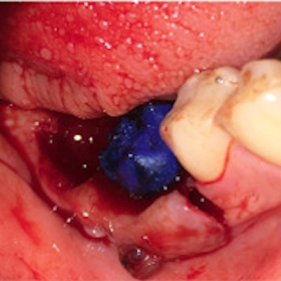
 Image caption and attribution: The tooth autotransplantation process: (A) Patient x-ray shows the upper third molar and destructed lower first molar. (B) Image of the recipient site before extraction. (C) Extractions and sockets debridement and disinfection. (D) The 3D-printed replica of the donor molar. (E) The 3D-printed replica is used to prepare the recipient site. (F) The third molar is transplanted to the new site. (G, H) The transplant site at follow-up. (I) X-rays two years later show no pathological signs. All images courtesy of Al-Khanati et al. Licensed under CC BY 4.0.
Selecting an ideal transplant candidate
The patient at the center of the case report was a 24-year-old woman who visited the oral surgery department complaining of an unpleasant smell and taste from her mouth and severe chewing dysfunction. She did not smoke and had an unremarkable medical history.
After completing a clinical and radiological exam, a clinician diagnosed the patient with destructive lower first and second molars on the right side with chronic infection. The molars required extraction, and cone-beam computed tomography (CBCT) imaging confirmed that the patient's upper right third molar could work as a donor tooth for autotransplantation.
The oral surgery department used the patient's CBCT scans to isolate and fabricate a replica of the third molar. The molar replica was then 3D printed and used to prepare the transplant site and check the fit before the procedure.
Tooth transplantation in under 20 seconds
On the day of the procedure, a clinician experienced in dental autotransplantation extracted the patient's first and second molars on the lower right side. They then used the 3D-printed replica tooth to prepare the recipient site.
When the site was ready for transplant, the clinician induced socket bleeding and instructed the assistant not to suction blood from the socket. The upper right third molar was then atraumatically extracted without the use of surgical burs.
The clinician moved the extracted third molar into the prepared lower first molar site in fewer than 20 seconds. The 3D-printed replica was then used to examine the occlusion, and the clinician completed the procedure by using silk sutures to immobilize the transplanted tooth.
The patient was instructed to eat a soft diet for one month and avoid touching the transplanted tooth as much as possible. She was prescribed antibiotics and a nonsteroidal anti-inflammatory drug, and she adhered well to the guidelines, the authors wrote.
Need to change transplant guidance?
Two years later, the patient returned to the clinic for periodontal probing, gingival index, mobility, percussion, occlusion, and pulp vitality testing. While the transplanted molar slightly rotated in the intervening time, the tooth was in good periodontal health and functional status, according to the authors.
More so, the tooth showed no discoloration and had a positive electric pulp vitality test. The patient said she was satisfied with the result.
"Immediate autotransplantation into an infected site can be successful if the recipient site is managed properly," the authors wrote.
The case demonstrates that molar transplantation can be successful in young patients who may not be the best candidates for implants. It also questions existing guidance around dental autotransplantation, including not performing molar transplantation in sites that have had an infection or without first performing root canal treatment.
"Hereby, we suggest changing the protocol and replacing it with one that limits [root canal treatment] to transplants that show clinical and/or radiographic signs of dental pulp necrosis," the authors wrote.