It was a year of interesting developments and eye-catching headlines in the world of digital dentistry. From the growth of artificial intelligence (AI) to radiology debates, there was plenty for dentists and dental team members to talk about in the world of digital dentistry in 2023.
With that in mind, let’s take a look back at the top five digital dentistry articles that DrBicuspid.com readers loved in 2023.
1. Dental x-rays in kids should be justified instead of routine
 Dr. Juan Yepes, MPH, MS, DrPh, of the department of pediatric dentistry at the Indiana University School of Dentistry and Riley Children's Hospital. Image courtesy of Dr. Yepes.
Though there is low value in taking routine dental x-rays in young children because it can lead to unneeded procedures, dentists should administer them when justified with factors, including caries risk status and exposure to fluoride, a pediatric dentist said.
Dr. Juan Yepes, MPH, MS, DrPh, of the department of pediatric dentistry at the Indiana University School of Dentistry and Riley Children's Hospital made these comments to DrBicuspid.com in response to an article published in Pediatrics that called for routine dental x-rays in children ages 3 to 6 to be targeted for de-implentation because they provided little value in diagnosing caries.
2. Actor Tom Hanks warns dental plan video is AI, not him
Oscar Award-winning actor Tom Hanks took to social media on October 1 to warn fans that a video promoting a dental plan included an unauthorized AI-generated image of Hanks.
On Instagram, Hanks wrote the following over the AI image, “"BEWARE!! There's a video out there promoting some dental plan with an AI version of me. I have nothing to do with it.”
3. Orthodontic wire travels to boy’s brain, causing a seizure
 (A) and (B) Coronal and sagittal view of the CT angiogram shows the wire penetrating through the skull floor of a 12-year-old boy. (C) Axial view of CT demonstrates the wire in the temporal lobe and the associated intraparenchymal hemorrhage. (D) Axial bone window CT shows the wire entering through the foramen ovale. Images courtesy of Morgan et al. Licensed by CC BY 4.0.
Imaging aided in the diagnosis of a 12-year-old boy in Texas who experienced a seizure after an orthodontic wire from his braces migrated into his temporal lobe.
After computed tomography (CT) scans and x-rays confirmed the metallic foreign object, which had traveled via the foramen ovale into the temporal lobe, as well as an associated intraparenchymal hemorrhage, the wire was removed without complications. The boy sustained no measurable damage to any structures within or around the foramen ovale, including the carotid artery, which would have been devastating, the authors wrote.
4. AAOMR: Stop shielding patients during dental imaging
The American Academy of Oral and Maxillofacial Radiology (AAOMR) announced that it no longer recommends patients’ reproductive organs and fetuses be covered during dental imaging, according to guidance published on August 1 in the Journal of the American Dental Association.
Additionally, the academy recommended discontinuing thyroid shielding during intraoral, panoramic, cephalometric, and cone-beam CT imaging, the authors wrote.
5. Imaging aids in diagnosis of man’s tooth with unusual root canal
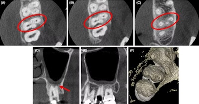 (A-C) Axial CBCT images in the cervical, middle, and apical region of the man’s molar. (D) The coronal section showed the apical split of roots and early periapical radiolucency in the palatal root (red arrow). (E) The sagittal section shows the gouging of the floor. Images courtesy of Marya et al. Licensed by CC BY 4.0.
Imaging aided in managing a man’s atypical root canal anatomy in which the distobuccal canal of his maxillary second molar was close to the palatal root canal with partially fused roots.
From the clinical and imaging findings, clinicians diagnosed the man with pulpal necrosis with symptomatic apical periodontitis and then executed appropriate endodontic treatment, the authors wrote.
