A dental implant that migrated into the maxillary sinus of a 47-year-old man caused an acute kidney infection secondary to sinusitis. The case report was published on February 13 in BMC Oral Health.
After the man's implant was removed, the sinus drained, and he underwent antibiotic therapy, the patient’s sinusitis and kidney infection resolved and didn’t recur again within the eight-year follow-up, the authors wrote.
“These findings suggest a complex bidirectional association or shared pathophysiological basis between odontogenic infections and multiple systemic diseases, including pyelonephritis,” wrote the authors, led by Gang Cheng of the Zhejiang Provincial People’s Hospital, Hangzhou Medical College, China.
A 47-year-old man with urinary problems, fever
In February 2016, the patient went to a nephrology outpatient clinic because of urinary frequency, urgency, and pain on urination for three days and a fever. His medical history was unremarkable, other than getting a dental implant in June 2015.
A physical exam and lab results revealed the man had acute pyelonephritis. He was treated with standard antibiotics for six months, but his condition continued.
His fever persisted, and he developed nasal congestion and a runny nose. It was revealed that he had an overflow of pus around his dental implant, and his sinuses were inflamed. The doctors suspected odontogenic maxillary sinusitis, the authors wrote.
 Figure 1: Dental implant proximal and distal mesial surfaces bonded to neighboring teeth with resin.Images and captions courtesy of Cheng et al. Licensed under CC BY-NC-ND 4.0.
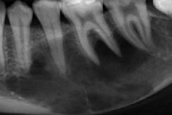
A cone-beam computed tomography (CBCT) scan of his mouth revealed that the implant was not well integrated with the bone. There was also chronic inflammation in the maxillary sinus. The next day, the defective implant was removed.
 Figure 2 (a-c): Cone-beam computed tomography (CBCT) scans taken at the initial consultation showed partial entry of the implant into the maxillary sinus, incomplete
osseointegration, inflammation in the maxillary sinus, and the implant crown
connecting with the neighboring tooth without a gap, (a) coronal view, (b) sagittal view, and (c) axial view.
A week later, the patient reported having a headache and a runny nose, and there was still pus in his mouth. He underwent a panoramic x-ray, which revealed a foreign body in the sinus that ended up being another dental implant. After using imaging to localize the second implant, it was surgically removed. Also, the patient underwent a sinus lift, they wrote.
 Figure 3: (a) Panoramic slice view after removal of the visible implant in the mouth
showing the presence of another implant in the maxillary sinus.
 Figure 4: (a) Bone window of approximately 1.5 cm x 1.5 cm buccal to the lateral wall of the maxillary sinus. (b) Removal of the lagging implant from the bone window. (c) Collagen membrane covering the bone graft to complete the maxillary sinus lift.
About nine months after the procedures, his sinus area returned to normal. Additionally, a new implant was placed.
 Figure 5: (a) Panoramic film taken after removal of the implant in the maxillary sinus for the maxillary sinus lift.
The patient was followed for eight years during which he maintained good overall health and did not experience a recurrence of acute pyelonephritis, the authors wrote.
Migrating dental implants
Whether clinicians should remove displaced implants from the maxillary sinus remains somewhat controversial, according to the case report.
The report highlights that a migrated implant can serve as a source of infection, which can spread to the bloodstream and to distant organs, such as the kidneys. Therefore, they should be removed promptly, they wrote.
“Postoperative maxillary sinusitis resolved, and no recurrence of pyelonephritis was observed during an 8-year follow-up, supporting a potential causal link as a distant source of infection,” Cheng and colleagues wrote.