After surviving a gunshot wound to the face, patient John Doe spent years seeking reconstructive care but was repeatedly turned away due to extensive maxillofacial trauma and bone loss. Nearly seven years later, he came to Dr. Safa Tahmasebi, MS, lead prosthodontist at the ClearChoice Dental Implant Center in Jackson, FL, and a faculty instructor with the Pikos Institute.
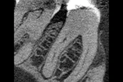
Recognizing the case’s complexity, Tahmasebi began with a high-resolution cone-beam computed tomography scan to assess residual bone and anatomical structures. The imaging revealed limited yet viable bone volume -- enough to anchor a customized full-arch implant solution. Using digital 3D modeling and surgical mirroring, Tahmasebi developed a precise treatment plan that guided implant placement and prosthetic design, allowing for complete oral rehabilitation and restoration of both function and aesthetics.
Below is an exclusive interview with Dr. Tahmasebi about the case, the challenges it presented, and how he overcame them to restore a smile. Please refer to the figures below as well for additional details.
Podcast takeaways
- The patient faced significant challenges due to a gunshot wound to the face.
- Innovative technology allows for 3D printing of patient anatomy for surgical planning.
- Understanding the emotional state of patients is crucial for effective treatment.
- The timeline for treatment can vary, but it typically spans several months.
- Patient commitment is essential for successful outcomes in complex cases.
- Not all dental procedures are suitable for every patient; careful planning is necessary.
- The emotional impact of dental rehabilitation can significantly change a patient's life.
- Collaboration with other specialists is often necessary for complex cases.
- Staying within the known aspects of dentistry is safer for practitioners.
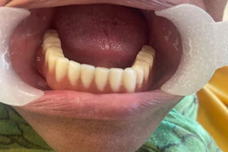
 Figure 1. Before (top image) and after (bottom image) of the reconstruction performed by Dr. Safa Tahmasebi, MS.All images courtesy of Dr. Safa Tahmasebi, MS.
 Figure 2: The man's upper jaw prior to reconstructive surgery.
 Figure 3: A forensic bone model demonstrates the bullet's trajectory and resulting damage to the man's mouth.
 Figure 4: John Doe's existing teeth were extracted and replaced with a maxillary full-arch implant.
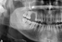
 Figure 5: Before and after panoramic x-ray of John Doe's teeth.
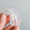
 Figure 6. The maxillary full-arch dental implant.
 Figure 7: 3D dental model of the maxillary full-arch implant.
 Figure 8: There was limited but viable enough bone to support a full-arch implant reconstruction.
 Figure 9: An anatomical model demonstrates how Dr. Tahmasebi reconstructed John Doe's maxilla.