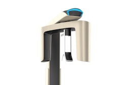
Detecting periapical lesions may vary depending on the imaging method, with three-dimensional cone-beam computed tomography (CBCT) providing higher detection rates. This review was recently published in the International Endodontic Journal.
However, using a combination of two-dimensional imaging techniques may yield results like those of CBCT, the authors wrote.
“Although imaging modality influences prevalence estimates, improved detection does not rely exclusively on three-dimensional imaging,” wrote the authors, led by Juan J. Segura-Egea of the University of Sevilla School of Dentistry in Spain (Int Endod J, July 30, 2026).
The goal of the review was to estimate the prevalence of periapical lesions in root-filled teeth and examine how different imaging methods influenced prevalence estimates. Researchers searched electronic databases through February 2026 and included studies that evaluated endodontically treated permanent teeth using CBCT, periapical radiography, or panoramic radiography, they wrote.
Eligible cross-sectional and cohort studies reported prevalence data at either the patient level, tooth level, or both. All 57 included studies had a cross-sectional design and were conducted across Europe, Asia, Africa, and South America in settings such as dental school clinics, university hospitals, private practices, and population-based samples. In total, the review analyzed data from 64,026 root-filled teeth and 35,569 patients.
The pooled prevalence of periapical lesions was 41.3% (95% confidence interval [CI], 38.2% to 44.6%) at the tooth level and 20.5% (95% CI, 9.3% to 39.4%) at the patient level. Furthermore, studies using CBCT consistently reported higher detection rates than most two-dimensional imaging techniques, they wrote.
However, combining panoramic and periapical radiographs produced prevalence estimates comparable to CBCT, whereas either method alone detected significantly fewer lesions. Considerable variability was observed across studies (I² = 98% to 99%), and the Egger's test (p = 0.048) suggested possible publication bias. Overall, most studies had a moderate risk of bias, and the certainty of the evidence was rated as low to very low.
Nevertheless, the study had limitations. Most studies relied on clinical or convenience samples from dental schools, hospitals, or radiographic databases, which may limit how accurately the findings represent the broader population, the authors added.
“The true burden of post-treatment apical pathology may be substantially underestimated when assessed using two-dimensional imaging alone,” Segura-Egea and colleagues concluded.