Researchers have developed a new x-ray detector material that produces clear dental images at radiation doses far below those used in commercial systems, according to a study published in National Science Review.
The technology addresses a persistent trade-off in radiography: Sharper images typically require higher radiation doses, while dose reduction tends to introduce visible noise, particularly problematic in low-dose scenarios such as pediatric exams, repeat imaging, or population-level screening.
The problem with thick detector films
The research team, working with Science China Press, identified that noise at low doses isn't solely a matter of fewer photons reaching the detector. X-ray photons arrive with natural statistical variation, and in a detector film that lacks uniformity through its thickness, absorption at different depths generates unevenly collected signals. That unevenness compounds the natural fluctuation into the grainy noise visible in low-dose images.
A new fabrication approach
To solve this, the team developed a technique called LPGA (liquid-phase growth and annealing), which stabilizes the liquid-phase environment during the formation of thick perovskite detector films. Rather than allowing temperature and composition gradients to develop unevenly during growth, LPGA produces a film that is uniform not just at the surface but throughout its full depth.
In testing, this improved internal consistency translated directly into lower image noise, a more stable x-ray response, and reliable image quality even under ultralow-dose conditions.
What LPGA means for dental imaging
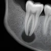
In a dental imaging demonstration, the LPGA detector clearly resolved crown and root structures at a dose substantially lower than that of a commercial dental x-ray system used for comparison. Additional tests on objects including a bat skeleton, a walnut, and an orange slice showed the detector preserved fine structural detail across varied sample types.
The authors note that the gains came entirely from the detector material itself, not from software-based noise reduction or improved electronics, suggesting a materials-science route to lower-dose imaging that could complement existing digital sensor and AI-analysis advances already reshaping dental radiography.
While still in the research stage, the findings point toward a potential new generation of intraoral and imaging sensors capable of maintaining diagnostic image quality at meaningfully reduced radiation exposure -- a development that could be particularly relevant for pediatric patients and practices performing frequent radiographic monitoring.