Deep subgingival decay can turn a seemingly routine posterior restoration into a complex clinical challenge. When caries extends below the cementoenamel junction, clinicians must manage multiple variables at once: limited access, reduced visibility, fluid contamination, tissue interference, matrix adaptation, and the need to preserve as much healthy tooth structure as possible.
In these cases, adhesive dentistry can provide a conservative and predictable restorative pathway when paired with careful isolation, magnification, and proper technique. The dental microscope further supports this approach by improving visualization of the subgingival area, helping the clinician evaluate the extent of decay, manage the gingival margin, and confirm matrix adaptation before restorative placement.
The following case demonstrates a technique for managing deep subgingival interproximal decay using magnification, rubber dam isolation, caries detection, selective enamel etching, immediate dentin sealing, resin coating, sectional matrix placement, and incremental composite placement.
Adhesive dentistry in deep subgingival cases
 Dr. Jorge F. Zapata.
Modern adhesive dentistry is built around the principle of preserving healthy tooth structure while creating a reliable bond to enamel and dentin. The resin coating concept was first proposed in Japan in the early 1990s by researchers, including Dr. Shigehisa Inokoshi and Dr. Toru Nikaido. It later became closely associated with adhesive dentistry concepts such as immediate dentin sealing and resin coating. Two important concepts in this approach are immediate dentin sealing (IDS) and the resin coating technique (RCT).
IDS involves applying an adhesive system to freshly cut dentin before final restorative placement. This technique helps seal exposed dentinal tubules, reduce the risk of postoperative sensitivity, and create a protected bonding surface.
RCT builds on IDS by placing a thin layer of flowable resin composite over the sealed dentin. This resin coating helps protect the hybridized dentin surface, isolate the pulpal floor, and create a more favorable restorative foundation. In many cases, a 0.5-mm to 1-mm layer of flowable composite is sufficient to protect the sealed dentin and help reduce irritation from external stimuli.
Together, IDS and RCT can be used in both direct and indirect restorative procedures. They also can be helpful in endodontically treated teeth by helping seal the coronal aspect of the restoration and reducing the potential for leakage beneath the definitive restoration.
Why subgingival interproximal decay is challenging
Subgingival decay occurs at or below the cementoenamel junction and may be associated with recurrent caries around an existing restoration, poor plaque control, restorative overhangs, margin breakdown, or systemic and behavioral factors that increase caries risk.
Interproximal subgingival lesions are especially difficult because the clinician must remove decay thoroughly while protecting adjacent soft tissue and maintaining control of the restorative field. Access may be limited by the interproximal papilla. Visibility may be compromised by the depth and location of the lesion. Fluid control can be difficult, and achieving a stable, well-adapted matrix can be challenging when the gingival margin is deep.
In many cases, treatment options may include crown lengthening; miniflap access; deep margin elevation; electrosurgery; laser-assisted soft-tissue management; orthodontic intervention; or conservative methods using retraction cord, wedges, Teflon tape, rubber dam isolation, and tissue compression. The appropriate approach depends on the location and extent of decay, periodontal considerations, restorative goals, and the clinician’s ability to achieve access and isolation.
The role of the dental microscope
Magnification and illumination can be especially valuable when treating deep subgingival decay. The dental microscope allows the clinician to evaluate the vertical and horizontal extent of the lesion, visualize the relationship between the gingival tissue and the subgingival wall, inspect the shape of the gingival floor, and confirm whether caries has been fully removed.
The microscope also supports more controlled matrix placement. With improved visualization, the clinician can better assess whether the sectional matrix is properly seated and adapted at the gingival margin before composite placement begins. This is critical, because even a small gap in the matrix can lead to overhangs, open margins, or excess restorative material in a difficult-to-finish area.
Case presentation
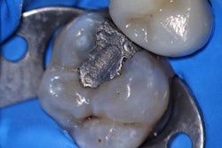
A patient presented with sensitivity in the upper left quadrant, particularly in response to cold. Radiographs revealed deep subgingival decay beneath an existing resin restoration on the distal aspect of tooth #13. Thermal testing was within normal limits, and the diagnosis was reversible pulpitis.
The treatment objective was to remove the defective restoration and recurrent decay, manage the deep subgingival margin, maintain isolation, and restore the tooth using an adhesive composite technique.
Clinical procedure
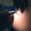
After local anesthesia was administered, a rubber dam was placed to isolate the tooth and improve moisture control. Isolation is essential in adhesive procedures, particularly when the restorative margin extends subgingivally and the contamination risk is increased.
The existing restoration was removed using a 330 carbide bur under 12 times magnification. Once the restoration was removed, surgical length round burs were used to remove soft, infected dentin and caries to the level of the gingival margin. Care was taken to avoid unnecessary trauma to the interproximal papilla while gaining access to the lesion.
Where tissue access limited visualization and restorative control, the interproximal papilla was carefully managed with the gingivectomy using electrosurgery or a laser. This allowed improved access to the gingival extent of the preparation while helping create the space needed for matrix placement and restorative finishing.
A caries indicator was then applied to confirm the removal of infected dentin. Any remaining stained, infected dentin was carefully removed. After caries excavation, a prophy jet cleaned residual biofilm from the tooth's surface. When bleeding occurred, a hemostatic agent was applied to regain control of the field before bonding.
Once the preparation was clean and controlled, attention turned to matrix placement. Because of the depth of the subgingival margin, a sectional matrix approximately 6 mm to 7 mm in height was selected. In some cases, the matrix may need to be customized on the buccal or lingual aspect to improve adaptation. A wedge was placed, and Teflon tape was used to help secure and adapt the matrix. A composite ring was then placed to stabilize the assembly.
With the matrix in place, the enamel was selectively etched using blue etch according to the manufacturer’s instructions. The adhesive protocol then incorporated IDS and RCT principles. A self-etch adhesive system was applied to the freshly prepared dentin. In this case, a Clearfil adhesive from Kuraray was selected because certain Clearfil adhesive systems incorporate MDPB, an antibacterial monomer designed to provide antibacterial activity within the adhesive layer.
After dentin sealing, a thin resin coating was placed to protect the sealed dentin and help isolate the pulpal floor. Composite resin was then placed incrementally, beginning at the gingival floor. Each increment was carefully adapted and light-cured before the next layer was placed. Incremental placement helps improve control in a deep preparation and may help reduce polymerization stress by managing the C-factor, or the ratio of bonded to unbonded surfaces.
To improve light access, the wings of the matrix were opened, allowing the restoration to be cured from the buccal, lingual, and occlusal aspects. After initial curing, the matrix was removed, and any remaining areas were light-cured as needed.
The restoration was then finished and polished to establish appropriate contour, function, and aesthetics.
Discussion
This case illustrates how adhesive dentistry can be applied in a challenging subgingival restorative scenario when the clinician is able to establish visibility, isolation, access, and matrix control.
The dental microscope played an important role throughout the procedure. Enhanced magnification and illumination supported caries removal, tissue management, evaluation of the gingival floor, and verification of matrix adaptation. This level of visibility is especially valuable when working in deep interproximal areas where direct vision is limited and restorative errors can be difficult to detect.
IDS and RCT also supported the restorative workflow by helping seal freshly cut dentin before composite placement. In a patient who presented with cold sensitivity and a diagnosis of reversible pulpitis, protecting the dentin and reducing exposure to external stimuli were important elements of the clinical approach.
The case also demonstrates the importance of using multiple adjuncts when necessary. Rubber dam isolation, caries indicator, prophy jet cleaning, hemostatic control, a customized sectional matrix, wedge placement, Teflon tape, and incremental composite placement all contributed to the ability to manage a difficult subgingival restoration conservatively.
Conclusion
Deep subgingival interproximal decay presents significant restorative challenges, particularly when visibility, isolation, tissue management, and matrix adaptation are compromised. By combining microscope-enhanced visualization with adhesive techniques such as IDS and RCT, clinicians can approach these cases with greater control and confidence.
In this case, magnification, careful caries removal, controlled soft-tissue management, selective enamel etching, dentin sealing, resin coating, and incremental composite placement helped support a conservative direct restorative solution for a tooth with deep recurrent subgingival decay.
Dr. Jorge F. Zapata is a general dentist in Ogden, UT, who has been practicing microscope dentistry in his private dental practice since 2005. He is a past executive board member and former treasurer of the Academy of Microscope Enhanced Dentistry. Zapata is an international lecturer who specializes in video and microphotography capture under the dental microscope. Zapata also lectures on the benefits of dental microscope usage in oral surgery, microphotography, and integration of cone-beam computed tomography. He serves as a key opinion leader for Kuraray Noritake Dental and Pascal Dental. He can be contacted at [email protected].
The comments and observations expressed herein do not necessarily reflect the opinions of DrBicuspid.com, nor should they be construed as an endorsement or admonishment of any particular idea, vendor, or organization.