CDOCS is an invaluable resource for dental professionals seeking to refine their surgical techniques. Among the platform's extensive library of educational content, five clinical tips shared with DrBicuspid.com readers this year resonated with readers. Each tip offers practical insights that can immediately improve patient outcomes.
Here's what makes these techniques essential viewing for any practitioner focused on oral surgery and implant dentistry.
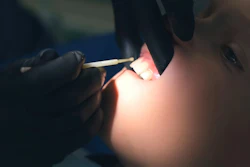
Sectioning teeth for oral surgery
Dr. Darin O'Bryan challenges conventional wisdom in his clinical tip on sectioning teeth for oral surgery. Rather than the traditional method of sectioning from the crown down, O'Bryan advocates starting at the furcation and working upward to the occlusal surface. This technique provides a crucial advantage: You know exactly how deep to go.
By creating an envelope flap that reveals the crest and furcation, clinicians can section teeth with precision while preserving critical bone, which is critical when hen planning for future implant placement. The technique also offers a better purchasing point for elevator placement, reducing the risk of coronal structure fractures that complicate extractions.
Mastering the double-socket membrane graft
When facing extensive socket defects with significant buccal and palatal bone loss, Dr. Farhad Boltchi's approach to double-socket membrane grafting provides an effective solution. This technique comprises a layered membrane strategy: a long-acting resorbable membrane (such as OSSIX Plus) serves as the primary layer, topped with a titanium-reinforced PTFE membrane as secondary protection.
The dual approach offers flexibility. Should the PTFE membrane become exposed or need early removal, the underlying resorbable membrane will protect the graft site. After five to six months of healing, clinicians can punch directly through the remaining OSSIX Plus membrane for implant placement, achieving ideal positioning in a reconstructed ridge.
A CBCT checklist for dental implant precision
The late Dr. Ross Enfinger's three-point checklist for dental implants and cone-beam computed tomography imaging is an essential protocol for virtual implant planning. Enfinger recommends using 360° viewing sliders to verify clearance of vital anatomic structures.
Next, clinicians should measure the distance from the implant platform to the gingival margin, ensuring approximately 3 mm of depth for proper restorative emergence. Finally, sleeve position verification confirms no complications with adjacent teeth or soft tissue. Enfinger's tip for improved visualization -- turning off the CT scan and virtual restoration views -- visualizes the relationship between the adjacent teeth and the sleeve position with greater clarity.
Guided bone profiling
For practitioners desiring to optimize bone architecture before placing an implant, this tip on guided bone profiling provides step-by-step procedural details. This approach allows precise control over ridge development, creating ideal implant sites through strategic bone augmentation.
Repositioning a luxated tooth
Dental trauma requires swift action. Dr. Doug Smail's guidance on repositioning a luxated tooth equips practitioners with the knowledge and confidence to handle these urgent situations effectively, preserving teeth that might otherwise be lost.
Why these tips matter
What sets these CDOCS clinical tips apart is their practical, immediately applicable nature. Each video features counsel from experienced clinicians sharing hard-won insights from years of practice. Whether you're refining established techniques or adding new procedures to your repertoire, these five resources represent essential viewing for modern dental practice.
Visit CDOCS.com to access these tips and explore their complete library of clinical education resources.