Dr. Daniel Vasquez.
In today’s digital dentistry, delivering restorations that are both high quality and efficient is no longer a wish, it’s an expectation. By combining the CEREC CAD/CAM ecosystem with Katana Zirconia One for Implant, clinicians can achieve aesthetic, durable, implant-supported crowns in a single appointment.
This case demonstrates a streamlined workflow using CEREC Primescan, CEREC software, and Primemill together with Katana Zirconia One for Implant. The result is a predictable, patient-friendly restoration that maintains clinical excellence while reducing chairtime and eliminating temporaries.
Case overview: Same-day restoration of an implant-supported crown
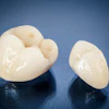
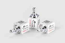
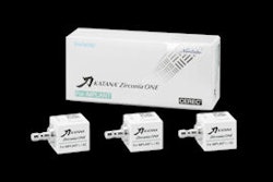
The workflow begins with the Katana Zirconia One for Implant block system (Figure 1), which includes three preshaded blocks and a demonstration restoration for reference.
 Figure 1: The Katana Zirconia One for Implant block system. All images courtesy of Kuraray America Inc.Kuraray America Inc.
Clinically, the case proceeded with an intraoral view of the healing abutment (Figure 2). After removal, a ScanPost was placed and scanned using CEREC Primescan (Figure 3), capturing a full-arch digital impression with precise scan body positioning and soft-tissue contours.
 Figure 2: The healing abutment in place.
 Figure 3: ScanPost placement for digital impression.
The definitive crown was then designed in CEREC software (Figure 4) using virtual articulation and occlusal analysis (Figure 5) and verified in a full-arch digital view for occlusal harmony (Figure 6). Milling was completed using CEREC Primemill with a Katana Zirconia One for Implant block, producing excellent marginal adaptation and lifelike translucency (Figure 7).
 Figure 4: Digital crown design in CEREC software.
 Figure 5: Virtual articulation and occlusal analysis.
 Figure 6: Full-arch digital verification.
 Figure 7: Final milled implant crown design from Katana Zirconia One block (Kuraray).
Surface treatment and cementation protocol
Surface preparation included sandblasting the intaglio of the zirconia crown and the titanium base with 50 µm aluminum oxide (Figures 8a and 8b) to increase micromechanical retention.
 Figure 8a: Sandblasting the Ti-base.
 Figure 8b: Sandblasting intaglio of the implant crown.
 Figure 9: Application of Clearfil Ceramic Primer Plus.
The crown was then luted to the titanium base using Panavia V5 Opaque (Figure 10), a dual-cure, self-etch resin cement also containing MDP chemistry for enhanced, reliable bonding. Ceramic primer was reapplied prior to final seating, and the interface was light-cured to optimize bond strength (Figure 11).
 Figure 10: Cementation with Panavia V5 Opaque.
 Figure 11: Light-curing of bonded interface.
Final seating and outcome
The hybrid crown was then tried in to verify occlusion and proximal contacts. (Figure 12). Once confirmed, the screw-access channel was sealed using Clearfil Majesty ES-2 composite to maintain aesthetics and function (Figure 13).
 Figure 12: The screw-access channel with crown attached.
 Figure 13: The screw-access channel was sealed using Clearfil Majesty ES-2 composite to maintain aesthetics and function.
A final radiograph (Figure 14) confirmed ideal seating, emergence profile, and a harmonious relationship with adjacent structures, completing the entire process in a single patient visit.
 Figure 14: Final radiograph showing seated restoration.
Why it works
The strength of this workflow lies in its integration with other systems. Each step -- from scanning to design, milling, and cementation -- is digitally driven and optimized for predictable, same-day delivery. Katana Zirconia One for Implant provides high strength and natural aesthetics while the MDP-based bonding system ensures robust, durable adhesion to the titanium base.
Because the block is preshaded, minimal staining or post-processing is needed, reducing both time and complexity. Together, these elements create a functional, aesthetic implant restoration without laboratory delays or provisional restorations.
Conclusion
Digital workflows are now a standard of care in implant dentistry. With the right combination of tools and materials, clinicians can confidently deliver implant-supported crowns that are strong, beautiful, and completed in just one appointment. The CEREC and Katana Zirconia One for Implant workflow makes that possible.
Dr. Daniel Vasquez has been practicing dentistry in San Diego since 1999, with a dedicated focus on aesthetic and digital dentistry. After acquiring his first CEREC system in 2008, Dr. Vasquez quickly became a recognized expert and opinion leader, collaborating as a beta tester with Kuraray Noritake, Dentsply Sirona Systems, TruAbutment Inc., and Vita North America. He regularly lectures nationally and internationally and mentors dentists worldwide in CAD/CAM dentistry.
The comments and observations expressed herein do not necessarily reflect the opinions of DrBicuspid.com, nor should they be construed as an endorsement or admonishment of any particular idea, vendor, or organization.